Infantile hepatic hemangioendothelioma: a clinicopathologic study in a Chinese population
- PMID: 20857525
- PMCID: PMC2945486
- DOI: 10.3748/wjg.v16.i36.4549
Infantile hepatic hemangioendothelioma: a clinicopathologic study in a Chinese population
Abstract
Aim: To investigate whether the clinicopathologic features of infantile hemangioendothelioma (IHE) of the liver in a Chinese population are similar to the features observed in other races.
Methods: The clinical data, radiological findings, histopathological changes and outcome of 12 cases of IHE diagnosed by the Department of Pathology, West China Hospital over the last 10 years were analyzed retrospectively. Immunohistochemical studies were carried out using antibodies against CD31, CD34, Factor VIII, cytokeratin 8 and cytokeratin 18.
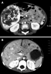
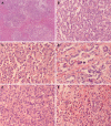
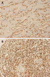
Results: The 12 patients were aged from fetal to 5 years (three males and nine females). The tumor was presented with different clinical manifestations, mainly as an asymptomatic, palpable, upper abdominal mass, except for the two fetuses who were detected antenatally by ultrasound. In one patient, this presentation was accompanied by an initial severe pneumothorax. No symptoms of congestive heart failure were present and neither congenital abnormalities nor vascular tumors in the skin or other organs were found. Laboratory abnormalities included leukocytosis (40%), anemia (60%), thrombocytosis (60%), hyperbilirubinemia (16.7%), abnormal liver function (50%) and increased α-fetoprotein (80%). Based on radiological findings and gross specimens, the tumor presented as a solitary lesion or a multifocal space-occupying lesion. The tumor size ranged from 5.0 cm × 3.5 cm × 2.0 cm to 13.8 cm × 9.0 cm × 7.7 cm, and the 0.2-1.1 cm nodules were diffusely distributed within the multifocal tumor. Seven cases were surgically resected, three cases underwent biopsy and the two fetuses were aborted. Histologically, nine cases were classified as type I and three as type II, presenting aggressive morphologic features, immature vessels, active mitosis and necrosis. An inflammatory component, predominantly eosinophilic granulocytes, sometimes obscured the nature of the tumor. Ten patients are alive after a follow-up of 1-9 years. Based on immunohistochemistry, the endothelial cells in all cases were positive for CD31, CD34 and polyclonal factor VIII antigen, whereas the scattered hyperplasia bile ducts were positive for cytokeratin 8 and cytokeratin 18.
Conclusion: The clinical manifestations of IHE are non-specific. There is no significant correlation between histological type and prognosis. The clinicopathologic features of IHE in Chinese patients may provide a clue to further evidence-based studies.
Figures


References
-
- Kunstadter RH. Hemangio-endothelioma of liver in infancy: case report and review of literature. Am J Dis Child. 1933;46:803–810.
-
- McLean RH, Moller JH, Warwick WJ, Satran L, Lucas RV Jr. Multinodular hemangiomatosis of the liver in infancy. Pediatrics. 1972;49:563–573. - PubMed
-
- Nazir Z, Pervez S. Malignant vascular tumors of liver in neonates. J Pediatr Surg. 2006;41:e49–e51. - PubMed
-
- Dehner LP, Ishak KG. Vascular tumors of the liver in infants and children. A study of 30 cases and review of the literature. Arch Pathol. 1971;92:101–111. - PubMed
-
- Finegold MJ, Egler RA, Goss JA, Guillerman RP, Karpen SJ, Krishnamurthy R, O'Mahony CA. Liver tumors: pediatric population. Liver Transpl. 2008;14:1545–1556. - PubMed
Publication types
MeSH terms
LinkOut - more resources
Full Text Sources
Medical
Research Materials

