

Clinicopathological features of gastric glomus tumor
- PMID: 20857536
- PMCID: PMC2945497
- DOI: 10.3748/wjg.v16.i36.4616
Clinicopathological features of gastric glomus tumor
Abstract
Aim: To study the clinicopathological features of gastric glomus tumor and review the related Chinese literature published in 1990-2010.
Methods: A case of gastric glomus tumor was reported. Clinicopathological findings in 56 cases of gastric glomus tumor were analyzed.
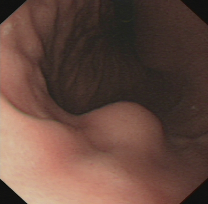
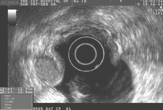
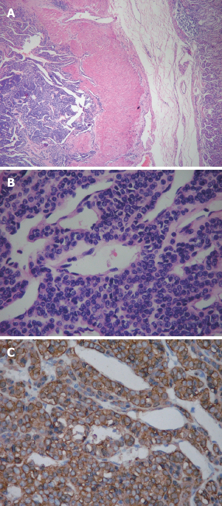
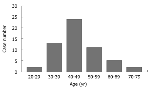
Results: Gastric glomus tumor was far more common in women than in men with a female to male ratio of 1.6:1. The median age of the patients was 45 years (range 28-79 years). The patients often complained of epigastric pain and bloody stool. The tumor was located in antrum of the stomach. The greatest diameter of the tumor was 0.8-11 cm. Histologically, the tumor was comprised of nests of glomus cells surrounding the capillaries. Glomus cells were small, uniform and round. Vimentin, smooth muscle actin and actin were expressed in the tumor. Other markers, including S-100 protein, CD34, CD117, desmin, CD56, synaptophysin, chromogranin A, neuron specific enolase and cytokeratin were all negative.
Conclusion: Gastric glomus tumor is a rare benign mesenchymal neoplasm. Its diagnosis depends on pathologic examination. Differential diagnosis includes gastrointestinal stromal tumor, paraganglioma and carcinoid tumor.
Figures
References
-
- Kay S, Callahan WP Jr, Murray MR, Randall HT, Stout AP. Glomus tumors of the stomach. Cancer. 1951;4:726–736. - PubMed
-
- Chou KC, Yang CW, Yen HH. Rare gastric glomus tumor causing upper gastrointestinal bleeding, with review of the endoscopic ultrasound features. Endoscopy. 2010;42 Suppl 2:E58–E59. - PubMed
-
- Chou HP, Tiu CM, Chen JD, Chou YH. Glomus tumor in the stomach. Abdom Imaging. 2010;35:390–392. - PubMed
MeSH terms
LinkOut - more resources
Full Text Sources
Medical
Research Materials
