. 2010 Sep 22;17(1):77.
doi: 10.1186/1423-0127-17-77.

High cell density and latent membrane protein 1 expression induce cleavage of the mixed lineage leukemia gene at 11q23 in nasopharyngeal carcinoma cell line

Affiliations

High cell density and latent membrane protein 1 expression induce cleavage of the mixed lineage leukemia gene at 11q23 in nasopharyngeal carcinoma cell line

Peter Han-Chung Yee et al. J Biomed Sci. .

Abstract

Background: Nasopharyngeal carcinoma (NPC) is commonly found in Southern China and South East Asia. Epstein-Barr virus (EBV) infection is well associated with NPC and has been implicated in its pathogenesis. Moreover, various chromosome rearrangements were reported in NPC. However, the underlying mechanism of chromosome rearrangement remains unclear. Furthermore, the relationship between EBV and chromosome rearrangement with respect to the pathogenesis of NPC has not been established. We hypothesize that during virus- or stress-induced apoptosis, chromosomes are initially cleaved at the base of the chromatin loop domain structure. Upon DNA repair, cell may survive with rearranged chromosomes.

Methods: In this study, cells were seeded at various densities to induce apoptosis. Genomic DNA extracted was processed for Southern hybridization. In order to investigate the role of EBV, especially the latent membrane protein 1 (LMP1), LMP1 gene was overexpressed in NPC cells and chromosome breaks were analyzed by inverse polymerase chain (IPCR) reaction.

Results: Southern analysis revealed that high cell density resulted in cleavage of the mixed lineage leukemia (MLL) gene within the breakpoint cluster region (bcr). This high cell density-induced cleavage was significantly reduced by caspase inhibitor, Z-DEVD-FMK. Similarly, IPCR analysis showed that LMP1 expression enhanced cleavage of the MLL bcr. Breakpoint analysis revealed that these breaks occurred within the matrix attachment region/scaffold attachment region (MAR/SAR).

Conclusions: Since MLL locates at 11q23, a common deletion site in NPC, our results suggest a possibility of stress- or virus-induced apoptosis in the initiation of chromosome rearrangements at 11q23. The breakpoint analysis results also support the role of chromatin structure in defining the site of chromosome rearrangement.


Figures
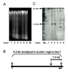
Figure 1
Figure 1
High cell density induces apoptosis and subsequent cleavage of the MLL bcr. (A) Ethidium bromide-stained agarose gel. SUNE1 (lanes 1-3) and HONE1 (lanes 4-6) seeded at cell number of 0.4 × 10⁵ (lanes 1 and 4), 2 × 10⁵ (lanes 2 and 5) and 4 × 10⁵ (lanes 3 and 6) were harvested for genomic DNA extraction after 4 days of growth. DNA was digested with BamH I and analyzed on 1% agarose gel. M represents the 1 kb DNA marker. (B) A schematic diagram illustrating the 8.3 kb MLL breakpoint cluster region (bcr). B represents the BamH I restriction site. Black box indicates the position of the DNA probe and down arrow shows the anticipated site of DNA cleavage. (C) Southern hybridization analysis. Southern hybridization was performed using the DNA probe shown in (B). Arrows labeled 8.3 kb and 1.5 kb show the positions of the intact and the cleaved MLL bcr respectively. MDig represents the DIG-labeled DNA marker (Roche, Penzberg, Germany).
Figure 2
Figure 2
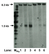
Caspase inhibitor reduces high cell density-induced MLL bcr cleavage. SUNE1 cell seeded at cell number of 0.4 × 10⁵ (lanes 1 and 4), 2 × 10⁵ (lanes 2 and 5) and 4 × 10⁵ (lanes 3 and 6) were allowed to grow for 4 days in the absence (lanes 1-3) or presence (lanes 4-6) of 50 μM caspase-3 inhibitor II (Z-DEVD-FMK). Extracted genomic DNA was processed for Southern hybridization as described in methods. Arrows labeled 8.3 kb and 1.5 kb show the positions of the intact and the cleaved MLL bcr respectively. MDig represents the DIG-labeled DNA marker.
Figure 3
Figure 3
Transfection of SUNE1 cell with LMP1 induces apoptotic cell death. SUNE1 cells were transiently transfected with pTracer vector (A and B) or LMP1 expression plasmid, pTracer-LMP1 (C and D). Cell morphology was monitored under bright-field microscopy (A and C) as well as dark field microscopy (B and D). Expression of the green fluorescence protein, GFP, is observed as green colored cells.
Figure 4
Figure 4
LMP1 expression induces cleavage of the MLL bcr. (A) Detection with anti-V5 antibody. SUNE1 cells were either transfected with vectors, pcDNA or pTracer (lanes 1 and 3); or LMP1 expression plasmids, pcDNA-LMP1 or pTracer-LMP1 (lanes 2 and 4). Cell lysate was analyzed on 10% SDS PAGE, and LMP1 expression was detected by anti-V5 antibody. (B) Detection with S12 anti-LMP1 antibody. SUNE1 cells were either transfected with vectors, pcDNA or pTracer (lanes 2 and 4); or LMP1 expression plasmids, pcDNA-LMP1 or pTracer-LMP1 (lanes 3 and 5). Cell lysate was analyzed on 10% SDS PAGE, and LMP1 expression was detected by S12 anti-LMP1 antibody. Lysate from the EBV-positive B95 cell line was included as positive control (lane 1). Arrows labeled 72 kDa and 63 kDa indicate the size of the expressed LMP1 (with V5 epitope and His-tag) and the endogenous LMP1 of B95-8 respectively. (C) Detection of MLL bcr cleavage by IPCR. SUNE1 cells transfected with vectors, pcDNA or pTracer (lanes 1 and 3); or LMP1 expression plasmids, pcDNA-LMP1 or pTracer-LMP1 (lanes 2 and 4) were collected for genomic DNA extraction. DNA was processed for nested IPCR as described in methods. Arrow labeled 2 kb indicates the position of the IPCR product of the intact MLL bcr. Arrows labeled 600 bp and 300 bp indicate the positions of the IPCR products of the cleaved MLL bcr. M1 and M2 represent the 1 kb and 100 bp DNA marker respectively.

References

    1. Fandi A, Altun M, Azli N, Armand JP, Cvitkovic E. Nasopharyngeal cancer: epidemiology, staging, and treatment. Semin Oncol. 1994;21:382–397. - PubMed
    1. Chien G, Yuen PW, Kwong D, Kwong YL. Comparative genomic hybridization analysis of nasopharygeal carcinoma: consistent patterns of genetic aberrations and clinicopathological correlations. Cancer Genet Cytogenet. 2001;126:63–67. doi: 10.1016/S0165-4608(00)00392-7. - DOI - PubMed
    1. Raab-Traub N. Epstein-Barr virus and nasopharyngeal carcinoma. Semin Cancer Biol. 1992;3:297–307. - PubMed
    1. Lin JC, Liao SK, Lee EH, Hung MS, Sayion Y, Chen HC, Kang CC, Huang LS, Cherng JM. Molecular events associated with epithelial to mesenchymal transition of nasopharyngeal carcinoma cells in the absence of Epstein-Barr virus genome. J Biomed Sci. 2009;16:105. doi: 10.1186/1423-0127-16-105. - DOI - PMC - PubMed
    1. Gan YY, Fones-Tan A, Chan SH, Gan LH. Epstein-Barr Viral Antigens Used in the Diagnosis of Nasopharyngeal Carcinoma. J Biomed Sci. 1996;3:159–169. doi: 10.1007/BF02253096. - DOI - PubMed

Publication types

MeSH terms

Substances