

Measurement of fetal biparietal diameter in owl monkeys (Aotus nancymaae)
Affiliations
- PMID: 20858355
- PMCID: PMC2949423
Item in Clipboard
Measurement of fetal biparietal diameter in owl monkeys (Aotus nancymaae)
J Am Assoc Lab Anim Sci.
2010 Sep.
Abstract
Owl monkeys are New World primates frequently used in biomedical research. Despite the historical difficulty of breeding owl monkeys in captivity, several productive owl monkey breeding colonies exist currently. The animals in the colony we describe here are not timed-pregnant, and determination of gestational age is an important factor in prenatal care. Gestational age of human fetuses is often determined by using transabdominal measurements of fetal biparietal diameter. The purpose of this study was to correlate biparietal diameter measurements with gestational age in owl monkeys. We found that biparietal diameter can be used to accurately predict gestational age in owl monkeys.
Figures

Ultrasound images of owl monkey fetuses during the second half of pregnancy. (A) Typical image showing the fetal head in a position for measuring gestational biparietal diameter (BPD). f, falx; t, thalamus. (B) Thorax. a, appendage; h, heart; p, placenta. (C) Abdomen. a, appendage; *, spine; s, stomach. Ultrasonography can be used to reliably assess fetal viability and morphology. During a typical exam, the following fetal structures are observed and evaluated: skull, heart, limbs, spine, stomach, and umbilical artery and vein (not shown). The placenta is evaluated for appropriate placement and echogenicity. BPD and fetal heart rate typically are measured also.

A graph of gestational biparietal diameter (BPD) of Aotus nancymaae fetuses. Measurements were made across the fetal head, with clear visualization of the thalamus, falx, and third ventricle at the level of the cavum septum pellucidi. BPD was determined at the widest point of the head circumference. Data were collected at random intervals throughout the pregnancies of 27 dams. Dams were not time-mated but were members of a breeding colony. Days prior to birth was calculated after birth by counting back to the date of the ultrasound exam and subtracted from the assumed term length of 133 d to obtain gestational age (that is, days of gestation). Linear regression was calculated by using the Marquardt–Levenberg (order 2) algorithm, with r2 = 0.94. Gestational age can be calculated as 12.11 + (43.66 × BPD) + [(BPD − 2.07)2 × 4.21].
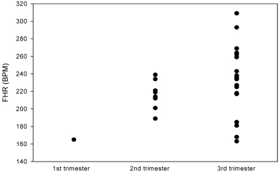
A scatter plot of fetal heart rate (FHR) as correlated to trimester. Although it varies considerably among animals, FHR is greater than 150 bpm in viable fetuses.
Similar articles
-
Ultrasonographic monitoring of a spontaneous abortion in an owl monkey (Aotus nancymaae).J Am Assoc Lab Anim Sci. 2007 Jul;46(4):74-6. J Am Assoc Lab Anim Sci. 2007. PMID: 17645301
-
Three novel neoplasms in Nancy Ma's owl monkeys (Aotus nancymaae).Vet Pathol. 2025 May;62(3):371-375. doi: 10.1177/03009858241300549. Epub 2024 Dec 18. Vet Pathol. 2025. PMID: 39692093 Free PMC article.
-
Uterine evaluation and gestation diagnosis in owl monkey (Aotus azarai infulatus) using the B mode ultrasound.J Med Primatol. 2006 Jun;35(3):123-30. doi: 10.1111/j.1600-0684.2006.00155.x. J Med Primatol. 2006. PMID: 16764669
-
The Owl Monkey (Aotus spp.) as an Animal Research Model-Part 1: Taxonomy, Geographic Distribution, Anatomy, and Behavior.J Med Primatol. 2025 Jun;54(3):e70022. doi: 10.1111/jmp.70022. J Med Primatol. 2025. PMID: 40275545 Review.
-
Transabdominal and transrectal ultrasonography of fetuses in Württemberg ewes: Correlation with gestational age.Anim Sci J. 2016 Feb;87(2):197-201. doi: 10.1111/asj.12421. Epub 2015 Jul 29. Anim Sci J. 2016. PMID: 26223772 Review.
Cited by
-
Ultrasonography of the neotropical primate female reproductive system.Front Vet Sci. 2024 Mar 8;10:1214509. doi: 10.3389/fvets.2023.1214509. eCollection 2023. Front Vet Sci. 2024. PMID: 38525406 Free PMC article. Review.
References
-
- Callen P. 1988. Ultrasonography in obstetrics and gynecology. Philadelphia (PA): WB Saunders Company
-
- Conrad SH, Sackett GP, Burbacher TM. 1989. Diagnosis of early pregnancy by ultrasound in Macaca fascicularis. J Med Primatol 18:143–154 - PubMed
-
- Corradini P, Recabarren M, Seron-Ferre M, Parraguez VH. 1998. Study of prenatal growth in the capuchin monkey (Cebus apella) by ultrasound. J Med Primatol 27:287–292 - PubMed
-
- Devonald KJ, Harewood WJ, Ellwood DA, Phippard AF. 1996. Fetal ultrasonography: normal biometric ranges in the baboon (Papio hamadryas). J Med Primatol 25:339–345 - PubMed
-
- Dixson AF. 1983. The owl monkey (Aotus trivirgatus), p 69–113 : Hearn JP. Reproduction in New World primates. Boston (MA): MTP Press Limited
Publication types
MeSH terms
Substances
Grants and funding
LinkOut - more resources
Full Text Sources