Review
. 2010 Dec 10:1364:90-102.
doi: 10.1016/j.brainres.2010.09.020. Epub 2010 Sep 19.

Anatomy of the kisspeptin neural network in mammals

Affiliations
Review

Anatomy of the kisspeptin neural network in mammals

Michael N Lehman et al. Brain Res. .

Abstract

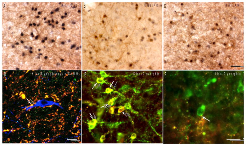
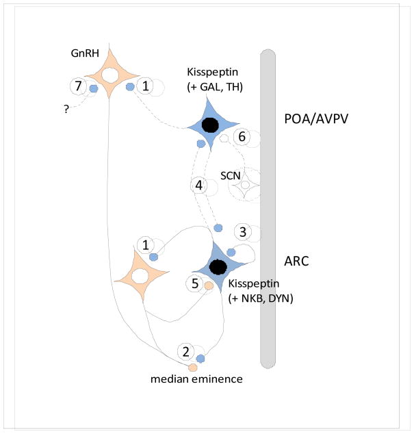
Kisspeptin has been recognized as a key regulator of GnRH secretion during puberty and adulthood, conveying the feedback influence of endogenous gonadal steroids onto the GnRH system. Understanding the functional roles of this peptide depends on knowledge of the anatomical framework in which it acts, including the location of kisspeptin-expressing cells in the brain and their connections. In this paper, we review current data on the anatomy of the kisspeptin neuronal network, including its colocalization with gonadal steroid hormone receptors, anatomical sites of interaction with the GnRH system, and recent evidence of neurochemical heterogeneity among different kisspeptin neuronal populations. Evidence to date suggests that kisspeptin cells in mammals comprise an interconnected network, with reciprocal connections both within and between separate cell populations, and with GnRH neurons. At the same time, there is more functional and anatomical heterogeneity in this system than originally thought, and many unanswered questions remain concerning anatomical relationships of kisspeptin neurons with other neuroendocrine and neural systems in the brain.


Figures

Fig. 1
Fig. 1
A–C: Colocalization of gonadal steroid receptors in kisspeptin neurons. Dual immunostained sections of sheep ARC showing high degree of colocalization of nuclear ER-α, PR and AR (blue-black) in kisspeptin cells (brown). Bar = 50 μm. D: Kisspeptin synaptic contacts onto a GnRH neuron. Confocal optical section (1 μm thick) of a triple-labeled section showing a terminal labelled with both kisspeptin (red) and synaptophysin (green) in direct contact with an ovine GnRH (blue) cell body (modified from Smith et al., 2008). Bar = 10 μm. E–F: Phenotypic heterogeneity between ARC and preoptic kisspeptin neurons. E: Colocalization of the endogenous opoid peptide, dynorphin (red), in kisspeptin neurons (arrows; green) of the sheep ARC. F: By contrast, kisspeptin neurons (green) in the sheep POA do not colocalize dynorphin even though they receive input from dynorphin-positive fibers (arrow; red). Bar = 20 μm. (modified from Goodman et al., 2007)
Fig. 2
Fig. 2. Schematic horizontal section (top=rostral, bottom=caudal) showing the POA/AVPV and ARC kisspeptin populations in the mammalian hypothalamus, and their potential sites of interactions with GnRH neurons
Virtually all ARC kisspeptin neurons co-express neurokinin B and dynorphin (Goodman et al., 2007; Navarro et al., 2009; Wakabayashi et al., 2010), while subsets of AVPV kisspeptin neurons in the preoptic region express either galanin (Vida et al., 2009) or tyrosine hydroxylase (Kauffman et al., 2007b). Connections (solid lines, published data; dotted lines, unpublished data) and sites of interactions between kisspeptin and GnRH systems include: 1) Direct projections from ARC and AVPV kisspeptin cells onto GnRH cell bodies (Clarkson and Herbison, 2006; Kinoshita et al., 2005; Krajewski et al., 2005; Lehman et al., 2010; Ramaswamy et al., 2008; Smith et al., 2008); 2) Inputs from ARC kisspeptin cells onto GnRH terminals in the median eminence (Burke, 2006; Krajewski et al., 2005; Lehman et al., 2010; Ramaswamy et al., 2008); 3) reciprocal connections among ARC kisspeptin cells that could be from the same or adjacent neurons (Burke, 2006; Foradori et al., 2002; Krajewski et al., 2010; Wakabayashi et al., 2010); 4) Projections from ARC kisspeptin neurons to POA kisspeptin cells in the sheep (Lehman, unpublished), and from AVPV kisspeptin neurons back to the ARC (Yeo and Herbison, 2009); and 5) projections from GnRH neurons back onto ARC kisspeptin cells (Ramaswamy et al., 2008). In addition, afferents to POA/AVPV kisspeptin cells from the suprachiasmatic nucleus (SCN) are indicated (6) (Vida et al., 2010), as well as the possibility that kisspeptin inputs to GnRH neurons in the POA or MBH may arise from other populations such as the DMH, BNST or medial amygdala (7).

References

    1. Adachi S, Yamada S, Takatsu Y, Matsui H, Kinoshita M, Takase K, Sugiura H, Ohtaki T, Matsumoto H, Uenoyama Y, Tsukamura H, Inoue K, Maeda KI. Involvement of Anteroventricular Periventricular Matastin/Kisspeptin Neurons in Estrogen Positive Feedback Action on Leutentizing Hormone Release in Female Rats. Journal of Reproduction and Development. 2007;53:367–378. - PubMed
    1. Adams VL, Goodman RL, Salm AK, Coolen LM, Karsch FJ, Lehman MN. Morphological Plasticity in the Neural Circuitry Responsible for Seasonal Breeding in the Ewe. Endocrinology. 2006;147:4843–4851. - PubMed
    1. Agnati LF, Zoli M, Strömberg I, Fuxe K. Intercellular communication in the brain: Wiring versus volume transmission. Neuroscience. 1995;69:711–726. - PubMed
    1. Ansel L, Bolborea M, Bentsen AH, Klosen P, Mikkelsen JD, Simonneaux V. Differential Regulation of Kiss1 Expression by Melatonin and Gonadal Hormones in Male and Female Syrian Hamsters. J Biol Rhythms. 2010;25:81–91. - PubMed
    1. Boehm U, Zou Z, Buck LB. Feedback Loops Link Odor and Pheromone Signaling with Reproduction. Cell. 2005;123:683–695. - PubMed

Publication types