Review
. 2010 Aug 30;62(11):1094-124.
doi: 10.1016/j.addr.2010.09.002. Epub 2010 Sep 19.

Development and applications of photo-triggered theranostic agents

Affiliations
Review

Development and applications of photo-triggered theranostic agents

Prakash Rai et al. Adv Drug Deliv Rev. .

Abstract

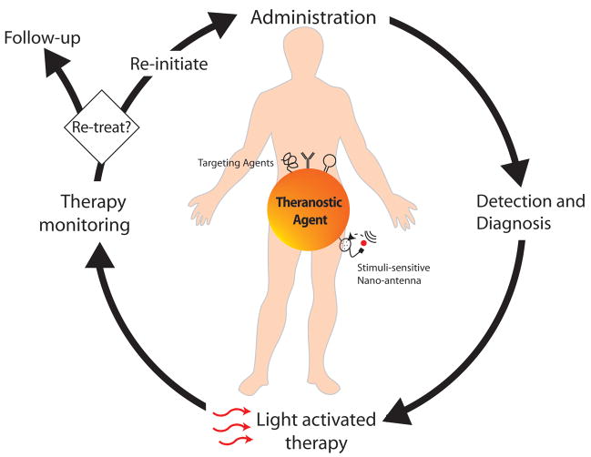
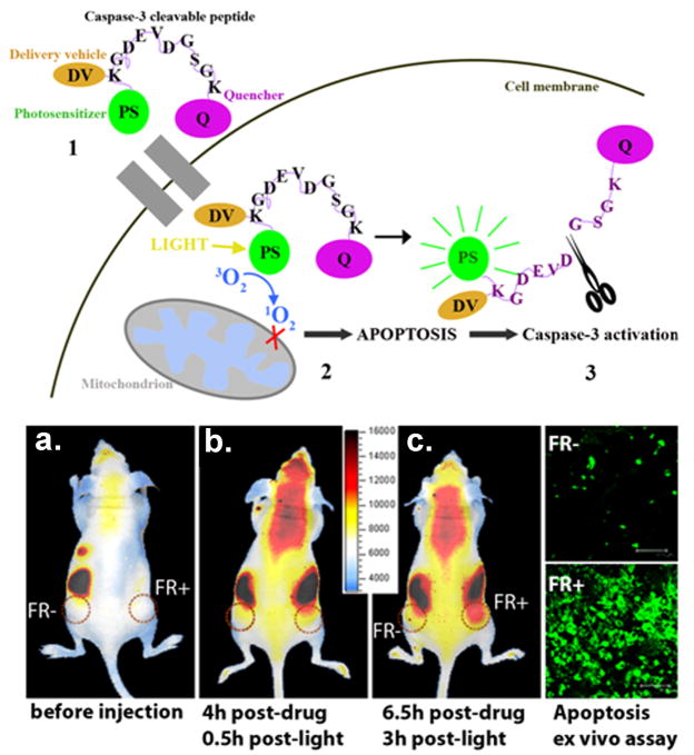
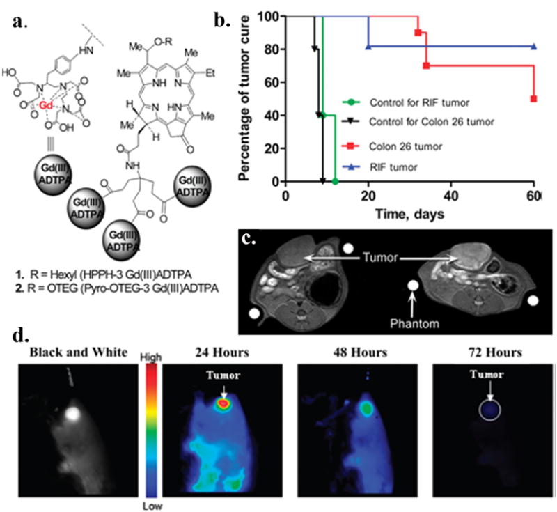
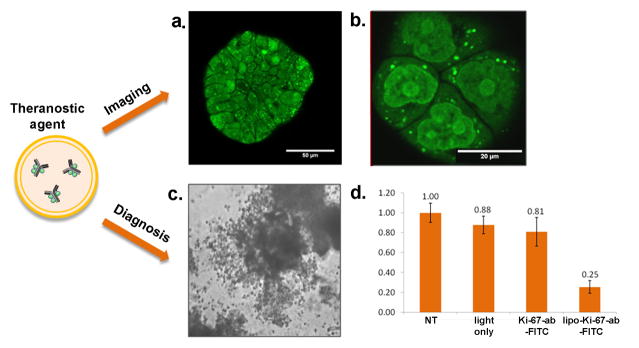
Theranostics, the fusion of therapy and diagnostics for optimizing efficacy and safety of therapeutic regimes, is a growing field that is paving the way towards the goal of personalized medicine for the benefit of patients. The use of light as a remote-activation mechanism for drug delivery has received increased attention due to its advantages in highly specific spatial and temporal control of compound release. Photo-triggered theranostic constructs could facilitate an entirely new category of clinical solutions which permit early recognition of the disease by enhancing contrast in various imaging modalities followed by the tailored guidance of therapy. Finally, such theranostic agents could aid imaging modalities in monitoring response to therapy. This article reviews recent developments in the use of light-triggered theranostic agents for simultaneous imaging and photoactivation of therapeutic agents. Specifically, we discuss recent developments in the use of theranostic agents for photodynamic-, photothermal- or photo-triggered chemotherapy for several diseases.


Figures

Figure 1
Figure 1
Role of photo-triggered theranostic agents at various stages of disease management. Following administration of a single, integrated theranostic agent, a clinician can diagnose disease, detect location of disease, deliver light at the disease locations for activating targeted therapy and following treatment, monitor response to therapy. At this stage, by monitoring the patients response to the therapy, the clinician can decide to either re-initiate treatment or if sufficient regression or cure of disease is observed,call the patient for a follow up visit.
Figure 2
Figure 2
Theranostic molecular beacons for targeted PDT and monitoring treatment response. The top panel is the schematic diagram of structure and function of a targeted PDT agent with a built-in apoptosis sensor: (1) This construct consists of PS, caspase 3 cleavable sequence, fluorescence quencher, and delivery vehicle; (2) The construct accumulates preferentially in cells overexpressing folate receptor, and once activated by light, the PS produces singlet oxygen that destroys the mitochondrial membrane and triggers apoptosis; (3) This leads to activation of caspase 3, which cleaves the peptide linker between the PS and the quencher, thus restoring the PS’s fluorescence and identifying those cells dying by apoptosis by NIR fluorescence imaging. The bottom left panel demonstrated in vivo induction and detection of apoptosis in a mouse bearing folate receptor positive (FR+, KB) cells) and folate receptor negative (FR-, HT 1080 cells) tumors after light treatment (Photodynamic Therapy = PDT, 90 J/cm2) using intravenously administered photoactivatable drug Pyro-K(folate)GDEVDGSGK (BHQ-3) (PFPB, 25 nmol) cleavable by Caspase-3. (a–c): Xenogen images of a mouse bearing FR- (left) and FR+ (right) tumors: a. before i.v. injection of PFPB or PDT; b. 0.5 hour after PDT (4 hours after drug injection); c. 3 hours after PDT (6.5 hours after drug injection). These images are showing a gradual increase in fluorescence in the FR+ compared to FR- tumor. Bottom right: Confocal images of the histology tissue slides of the corresponding FR+ and FR- tumors stained with Apoptag confirmed increased light-induced apoptosis in the FR+ tumor. Adapted from Steffalova et al. [42].
Figure 3
Figure 3
Tumor-Avid PS-Gd(III)DTPA for imaging and therapy. Conjugate shows potential for in vivo imaging (MR, Fluorescence) and PDT. a. Structure of Gd+3aminobenzyl DTPA conjugates of HPPH; b. In vivo PDT efficacy of HPPH-3Gd in C3H mice bearing RIF tumors and BALB/c mice bearing Colon 26 tumors. At an imaging/therapeutic dose of 10 umol/kg; c. Increase in signal intensity was seen in rat Ward Colon tumors (arrow) from preinjection (left) to postinjection (right, 24 hour postinjection) of HPPH-3Gd; d. Fluorescent images of HPPH-3Gd in BALB/c mice at 24, 48, and 72 hour. Adapted from Spernyak et al. [49].
Figure 4
Figure 4
Photoimmunoconjugate encapsulating liposomes (PICELS) for targeting and inactivation of the nuclear proliferation marker pKi-67. a. PICELS deliver pKi-67-FITC antibodies intracellular and can be imaged with confocal microscopy in ovarian cancer 3D-culture ascini. b. Imaging of monolayer cultures shows the nucleolar localization on the PICELS. c.72 hours after laser irradiation with 5 J/cm2 at 488 nm the 3D-acini have lost their spherical morphology and d. Following treatment with PICELS and light irradiation the 3D-acini show a significant decrease in the number of viable cells as measured by a live-dead assay. Based on work by Rahmanzadeh et al. [86].
Figure 5
Figure 5
Vascular targeted PDT with theranostic agents improves brain cancer therapy as confirmed by MRI a. Schematic representation of the multifunctional nanoparticles. The core of the nanoparticle was synthesized from polyacrylamide, which was embedded with PDT dyes (Photofrin) and/or imaging agents (magnetite/fluorochrome). Polyethylene glycol linker and a molecular address tag (F3 peptide) were attached to target these nanoparticles to cancer cells. b. Cytotoxicity induced by F3-tagged Photofrin-embedded nanoparticles and laser irradiation. MDA-435 cells were incubated 4 hours with nanoparticles with or without F3 tag and irradiated with 1,500 mW of laser for 5 minutes. The Photofrin-mediated cytotoxicity was then monitored by labeling cells with calcein-AM (green, live cells) and propidium iodide (dead, red cells). Bar, 20 μm. c. T2-weighted magnetic resonance images at day 8 after treatment from (C) a representative control i.c. 9L tumor and tumors treated with (D) laser light only, (E) i.v. administration of Photofrin plus laser light, and (F) nontargeted nanoparticles containing Photofrin plus laser light and (G) targeted nanoparticles containing Photofrin plus laser light.The image shown in (H) is from the same tumor shown in (G), which was treated with the F3-targeted nanoparticle preparation but at day 40 after treatment.The color diffusion maps overlaid on top ofT2-weighted images represent the apparent diffusion coefficient (ADC) distribution in each tumor slice shown. d. Kaplan-Meier survival plot for the i.c. 9L tumor groups. Survival curves for brain tumor animals: untreated, laser only, i.v. Photofrin + laser treated, nontargeted nanoparticles containing Photofrin + laser, and F3-targeted Photofrin-containing nanoparticles + laser treated. Adapted from Reddy et al. [100].
Figure 6
Figure 6
Gold nanorods, a theranostic agent, used for combined ultrasound and photoacoustic imaging and photothermal therapy. a. the TEM images of gold nanorods. b. Ultrasound and photoacoustic images of mouse tumor injected with gold nanorods. The tumor region is shown in white inset in the ultrasound image. The photoacoustic image shows higher contrast in the tumor area due to accumulation of gold nanorods. c. Thermal image of a subcutaneous tumor in nude mouse. During the PTT procedure approximately 25°C temperature rise was observed in the tumor. Adapted from Mallidi et al. [135].
Figure 7
Figure 7
Gold nanorods as theranostic agents for in-vivo X-ray CT imaging and PTT therapy. a and b show the feasibility of using nanorods as X-ray CT contrast agent. Clearly the images in b identify the location of the tumor marked by arrows. c and d showcase the feasibility of using nanorods as photothermal agents. d. shows the temperature rise in the tumor injected with gold nanorods during PTT. Finally, the in-vivo mouse survival studies indicate mice injected with PEGylated nanorods and undwernet NIR laser irradiation had greater survival rate than the control groups. Adapted from von Maltzahn et al. [164].
Figure 8
Figure 8
Photo-activatable theranostic agents for triggered release of drug. a. Use of light for gene delivery using the principle of photochemical internalization. Adapted from Nishiyama et al. [170] b. NIR light triggered release of drugs trapped in PLGA microspheres that also encapsulate hollow gold nanoshells. NIR light is absorbed by the HAuNSs and is converted to heat which triggers release of the encapsulated drug. Adapted from You et al. [171] c. Use of fluorescence optical imaging for highly selectively tumor imaging with an activatable fluorescence probe–antibody conjugate. The probe is nonfluorescent when outside the tumor cells. After internalization by endocytosis, the probe is accumulated in late endosomes or lysosomes, where the acidic pH activates the probe, making it highly fluorescent which is captured by optical imaging. Adapted from Urano et al. [172].
Figure 9
Figure 9
Enzyme activated theranostic prodrug for overcoming bacterial resistance a. Mechanism for the activation of β-LEAP. In resistant bacteria β-LEAP is cleaved by β-lactamase, releasing the quenched PS (blue balls) into an unquenched state (red balls). b. The released PS is activated by light irradiation, as shown in coculture of HFF-1 cells and strain 8179 (left panel) and HFF-1 cells alone (right panel). c. Inhibition profiles for selected strains of S. aureus with penicillin G for comparison are shown in. d. β-LEAP hydrolysis by selected strains of S. aureus leads to loss of viability in bacterial cells. Key for c and d: purple 29213, green 9307, gold 8150, red 8179, blue 8140 (d only). Based on work by Zheng et al. [212].
Figure 10
Figure 10
Theranostic agents for in vivo imaging and PDT of rheumatoid arthritis. a. Scheme illustrating the concepts involved in the design of the theranostic prodrug. Self-quenching of PS occurs when tethered to lysine backbone. After proteolytic cleavage and light activation, PS forms reactive oxygen species. b. Fluorescence of arthritic joints and non-arthritic joints pre (left image) and 4 h post i.v.administration (right image) of prodrug. Adapted from Gabriel et al. [230].

Similar articles

Cited by

  • Application of chitosan-based nanocarriers in tumor-targeted drug delivery.
    Ghaz-Jahanian MA, Abbaspour-Aghdam F, Anarjan N, Berenjian A, Jafarizadeh-Malmiri H. Ghaz-Jahanian MA, et al. Mol Biotechnol. 2015 Mar;57(3):201-18. doi: 10.1007/s12033-014-9816-3. Mol Biotechnol. 2015. PMID: 25385004 Review.
  • Diverse Applications of Nanomedicine.
    Pelaz B, Alexiou C, Alvarez-Puebla RA, Alves F, Andrews AM, Ashraf S, Balogh LP, Ballerini L, Bestetti A, Brendel C, Bosi S, Carril M, Chan WC, Chen C, Chen X, Chen X, Cheng Z, Cui D, Du J, Dullin C, Escudero A, Feliu N, Gao M, George M, Gogotsi Y, Grünweller A, Gu Z, Halas NJ, Hampp N, Hartmann RK, Hersam MC, Hunziker P, Jian J, Jiang X, Jungebluth P, Kadhiresan P, Kataoka K, Khademhosseini A, Kopeček J, Kotov NA, Krug HF, Lee DS, Lehr CM, Leong KW, Liang XJ, Ling Lim M, Liz-Marzán LM, Ma X, Macchiarini P, Meng H, Möhwald H, Mulvaney P, Nel AE, Nie S, Nordlander P, Okano T, Oliveira J, Park TH, Penner RM, Prato M, Puntes V, Rotello VM, Samarakoon A, Schaak RE, Shen Y, Sjöqvist S, Skirtach AG, Soliman MG, Stevens MM, Sung HW, Tang BZ, Tietze R, Udugama BN, VanEpps JS, Weil T, Weiss PS, Willner I, Wu Y, Yang L, Yue Z, Zhang Q, Zhang Q, Zhang XE, Zhao Y, Zhou X, Parak WJ. Pelaz B, et al. ACS Nano. 2017 Mar 28;11(3):2313-2381. doi: 10.1021/acsnano.6b06040. Epub 2017 Mar 14. ACS Nano. 2017. PMID: 28290206 Free PMC article. Review.
  • All-in-one processing of heterogeneous human cell grafts for gene and cell therapy.
    Lukianova-Hleb EY, Yvon ES, Shpall EJ, Lapotko DO. Lukianova-Hleb EY, et al. Mol Ther Methods Clin Dev. 2016 Mar 16;3:16012. doi: 10.1038/mtm.2016.12. eCollection 2016. Mol Ther Methods Clin Dev. 2016. PMID: 27006970 Free PMC article.
  • Light Sensitive Liposomes: A Novel Strategy for Targeted Drug Delivery.
    Aundhia C, Parmar G, Talele C, Talele D, Seth AK. Aundhia C, et al. Pharm Nanotechnol. 2025;13(1):41-54. doi: 10.2174/0122117385271651231228073850. Pharm Nanotechnol. 2025. PMID: 38279711 Review.
  • A supramolecular nanovehicle toward systematic, targeted cancer and tumor therapy.
    Liang R, You S, Ma L, Li C, Tian R, Wei M, Yan D, Yin M, Yang W, Evans DG, Duan X. Liang R, et al. Chem Sci. 2015 Oct 1;6(10):5511-5518. doi: 10.1039/c5sc00994d. Epub 2015 Jun 22. Chem Sci. 2015. PMID: 28717446 Free PMC article.

References

    1. Rising Health Costs a Global Problem, in, 2010.

    1. WORLD HEALTH STATISTICS 2010. World Health Organization; France: 2010. pp. 1–177.
    1. Rai AJ, Yee J, Fleisher M. Biomarkers in the era of personalized medicine - a multiplexed SNP assay using capillary electrophoresis for assessing drug metabolism capacity. Scand J Clin Lab Invest Suppl. 2010;242:15–18. - PubMed
    1. Blanchet KD. Redefining personalized medicine in the postgenomic era: developing bladder cancer therapeutics with proteomics. BJU Int. 2010;105:i–iii. - PubMed
    1. Francke U. On the bumpy road towards ‘personalized medicine’. EMBO Mol Med. 2010;2:1–2. - PMC - PubMed

Publication types