. 2010 Dec 1;19(23):4625-33.
doi: 10.1093/hmg/ddq393. Epub 2010 Sep 21.

Shox2 mediates Tbx5 activity by regulating Bmp4 in the pacemaker region of the developing heart

Affiliations

Shox2 mediates Tbx5 activity by regulating Bmp4 in the pacemaker region of the developing heart

Sandra Puskaric et al. Hum Mol Genet. .

Abstract

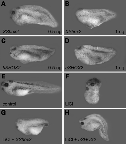
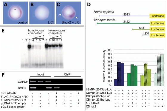
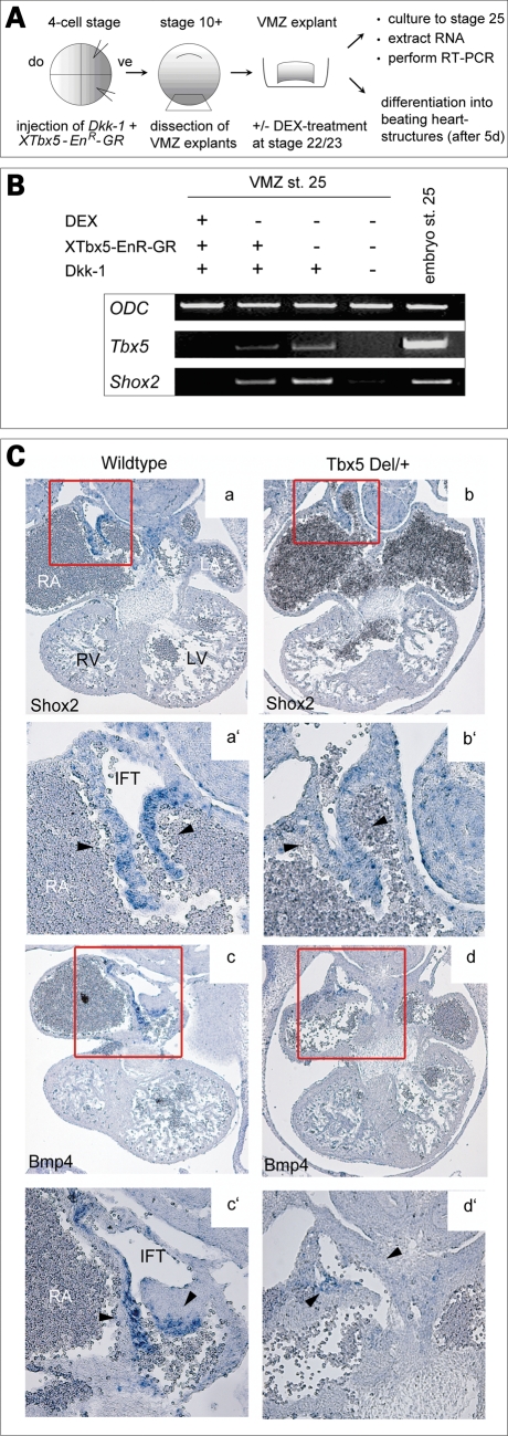
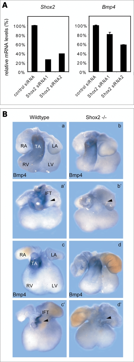
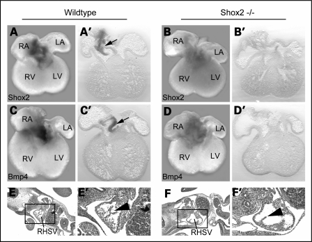
Heart formation requires a highly balanced network of transcriptional activation of genes. The homeodomain transcription factor, Shox2, is essential for the formation of the sinoatrial valves and for the development of the pacemaking system. The elucidation of molecular mechanisms underlying the development of pacemaker tissue has gained clinical interest as defects in its patterning can be related to atrial arrhythmias. We have analyzed putative targets of Shox2 and identified the Bmp4 gene as a direct target. Shox2 interacts directly with the Bmp4 promoter in chromatin immunoprecipitation assays and activates transcription in luciferase-reporter assays. In addition, ectopic expression of Shox2 in Xenopus embryos stimulates transcription of the Bmp4 gene, and silencing of Shox2 in cardiomyocytes leads to a reduction in the expression of Bmp4. In Tbx5(del/+) mice, a model for Holt-Oram syndrome, and Shox2(-/-) mice, we show that the T-box transcription factor Tbx5 is a regulator of Shox2 expression in the inflow tract and that Bmp4 is regulated by Shox2 in this compartment of the embryonic heart. In addition, we could show that Tbx5 acts cooperatively with Nkx2.5 to regulate the expression of Shox2 and Bmp4. This work establishes a link between Tbx5, Shox2 and Bmp4 in the pacemaker region of the developing heart and thus contributes to the unraveling of the intricate interplay between the heart-specific transcriptional machinery and developmental signaling pathways.


Figures

Figure 1.
Figure 1.
Ectopic Shox2 expression induces a ventralizing effect during early Xenopus development and rescues embryos partially dorsalized by LiCl. Lateral view of stage 36 embryos, radially injected at 4-cell stage with 0.5 ng (A, C) and 1 ng (B, D) Xenopus tropicalis Shox2 RNA (A, B) and human SHOX2a RNA (C, D). Shox2-injected embryos show a dose-dependent ventralizing effect. Uninjected stage 36 control embryo (E) and control embryo treated with 120 mM LiCl (F). Ventral injection of 1 ng Shox2 RNA rescues embryos partially dorsalized by LiCl (G, H).
Figure 2.
Figure 2.
Shox2 regulates the expression of Bmp4. (A) Bmp4 expression in uninjected embryos at stage 10.5 (n=35). Black arrowhead indicates the dorsal lip. (B) Bmp4 expression is reduced or absent in 87% of LiCl-treated embryos (n=31). (C) Spots of Bmp4 expression in 77% of previously Shox2-injected, LiCl-treated embryos (n=30). Shox2 RNA was injected diagonally into two blastomers of a 4-cell stage embryo. Bmp4 staining in two areas of the embryo (marked by white arrows) indicates an upregulation of Bmp4 expression corresponding to the Shox2 injection. (D) Shox2 increases the activity of the human and Xenopus laevis Bmp4 promoter upon co-transfection of Shox2 expression plasmids (1 µg) and the indicated Bmp4 reporter-constructs (1 µg) into Cos-7 cells. Data using HEK-293 cells are not shown. (E) Electrophoretic mobility shift assay of the GST–SHOX2 fusion protein on a conserved promoter oligonucleotide sequence (BMP4). An excess of non-radioactively labelled oligonucleotides (homologous competitor) reduces the DNA-binding ability of SHOX2, whereas excess of a random oligonucleotide sequence (heterologous competitor) does not affect DNA-binding. 1, free oligonucleotide; 2, GST alone; 3, GST–SHOX2; 4–7, 10-fold, 50-fold, 75-fold and 150-fold molar excess of homologous competitor; 8, GST–SHOX2; 9–12, 10-fold, 50-fold, 75-fold and 150-fold molar excess of heterologous competitor. (F) Chromatin immunoprecipitation (ChIP) assay. HEK-293 cells were co-transfected with the FLAG-SHOX2a expression or empty control vector and with either the hBMP4 2013-Luc reporter or empty control vector as indicated. Formaldehyde-crosslinked DNA was immunoprecipitated using an anti-FLAG antibody (α-FLAG-Ab) or no antibody, as a negative control. Precipitated DNA fragments (ChIP) and DNA from lysate before immunoprecipitation (Input) were subjected to PCR using primer sets amplifying the putative SHOX2-binding element in the BMP4 gene (lower panel) or GAPDH (upper panel) as a control.
Figure 3.
Figure 3.
Shox2 mediates Tbx5 expression to Bmp4 signaling in the developing heart. (A) Scheme of the ventral marginal zone (VMZ) experiment. Xenopus embryos were injected (vegetal dorsal) with 1 ng Dkk-1 mRNA and 0.2 ng of XTbx5 EnR-GR DNA into two blastomeres at the 4-cell stage and VMZ explants were dissected at early gastrula stage. Explants were cultured until stage 22/23 and then treated with 0.5 µM dexamethasone (DEX)-solution to activate the injected repressor-construct. RNA was extracted at stage 25 and analysed by RT–RCR for the presence of XTbx5 and XShox2. (B) RT–PCR shows that Dkk-1 induces Tbx5 and Shox2 expression in VMZ explants. XTbx5 EnR-GR abolishes Dkk-1 induced Tbx5 and Shox2 expression after DEX-treatment. Ornithine decarboxylase was used as a loading control. (C) Section in situ hybridization on E11.5 wild-type and Tbx5del/+ mouse hearts. Murine Shox2 is strongly expressed in the inflow tract of the developing heart in wild-type embryos (a), which is markedly decreased in Tbx5del/+ embryos (b). Murine Bmp4 expression overlaps with the Shox2 expression domain in wild-type hearts (c) and is almost absent in Tbx5del/+ embryos (d). RA, right atrium; LA, left atrium; RV, right ventricle; LV, left ventricle.
Figure 4.
Figure 4.
Shox2 deficiency impairs Bmp4 expression. (A) H10 cells were transfected with two different Shox2 siRNAs (Shox2 siRNA1 and Shox2 siRNA2) in parallel with a control siRNA. Expression levels of Shox2 (left panel) and Bmp4 (right panel) were assessed 24 h after transfection by qRT–PCR analysis. siRNA-mediated knock-down of Shox2 results in 19–42% reduction of Bmp4 mRNA levels. All results were normalized to Hprt1 (hypoxanthine phosphoribosyltransferase 1) mRNA values. (B) Whole-mount in situ hybridization on E11.5 (a, b) and E12.5 (c, d) wild-type and Shox2−/− mouse hearts using a Bmp4 RNA probe. Both, ventral (a–d) and dorsal (a′–d′) views are shown. Murine Bmp4 is strongly expressed in the truncus arteriosus (TA) (a, c) and in the inflow tract (a′, c′) of the developing heart in wild-type embryos. In the Shox2−/− mouse hearts, Bmp4 expression is still present in the truncus arteriosus (b, d) but completely absent in the IFT (b′, d′), where Shox2 and Bmp4 expression domains overlap in the wild-type heart. RA, right atrium; LA, left atrium; RV, right ventricle; LV, left ventricle.
Figure 5.
Figure 5.
Bmp4 expression is absent in the inflow tract (IFT) of Shox2−/− embryos. Whole-mount in situ hybridization with corresponding sections (50 µm) on E12.5 wild-type and Shox2−/− mouse hearts using Shox2 (A, B) and Bmp4 (C, D) RNA probes. Murine Shox2 (A, A′) and Bmp4 (C, C′) are expressed in the IFT of wild-type hearts (indicated by black arrows), but are completely absent in the IFT of Shox2−/− hearts (B, B′, D, D′). Note that IFT tissue is still present in Shox2−/− hearts (B′, D′). Histological sections of wild-type (E, E′) and Shox2−/− (F, F′) embryos at E10.5. Squares in E and F show the right horn of the sinus venosus that is magnified in E′ and F′. Shox2-deficient embryos develop IFT tissue (F, F′), even if the venous valve formation is abnormal (black arrowhead in F′). RA, right atrium; LA, left atrium; RV, right ventricle; LV, left ventricle; RHSV, right horn of the sinus venosus.

Similar articles

Cited by

References

    1. Bruneau B.G. The developmental genetics of congenital heart disease. Nature. 2008;451:943–948. - PubMed
    1. Boyett M.R., Honjo H., Kodama I. The sinoatrial node, a heterogeneous pacemaker structure. Cardiovasc. Res. 2000;47:658–687. - PubMed
    1. Christoffels V.M., Smits G.J., Kispert A., Moorman A.F. Development of the pacemaker tissues of the heart. Circ. Res. 2010;106:240–254. - PubMed
    1. Blaschke R.J., Hahurij N.D., Kuijper S., Just S., Wisse L.J., Deissler K., Maxelon T., Anastassiadis K., Spitzer J., Hardt S.E., et al. Targeted mutation reveals essential functions of the homeodomain transcription factor Shox2 in sinoatrial and pacemaking development. Circulation. 2007;115:1830–1838. - PubMed
    1. Espinoza-Lewis R.A., Yu L., He F., Liu H., Tang R., Shi J., Sun X., Martin J.F., Wang D., Yang J., et al. Shox2 is essential for the differentiation of cardiac pacemaker cells by repressing Nkx2–5. Dev. Biol. 2009;327:376–385. - PMC - PubMed

Publication types

MeSH terms