. 2010 Sep 21;3(140):ra67.
doi: 10.1126/scisignal.2001083.

Synthetic lethal screen of an EGFR-centered network to improve targeted therapies

Affiliations

Synthetic lethal screen of an EGFR-centered network to improve targeted therapies

Igor Astsaturov et al. Sci Signal. .

Abstract

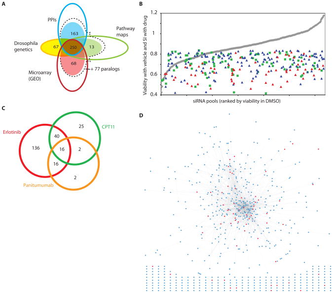
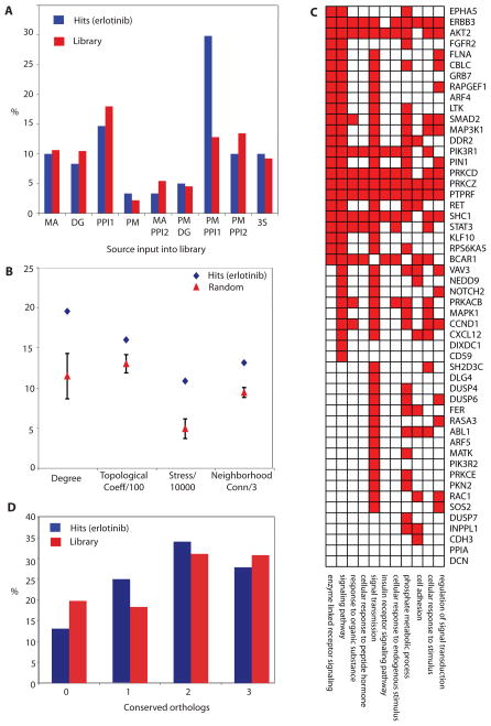
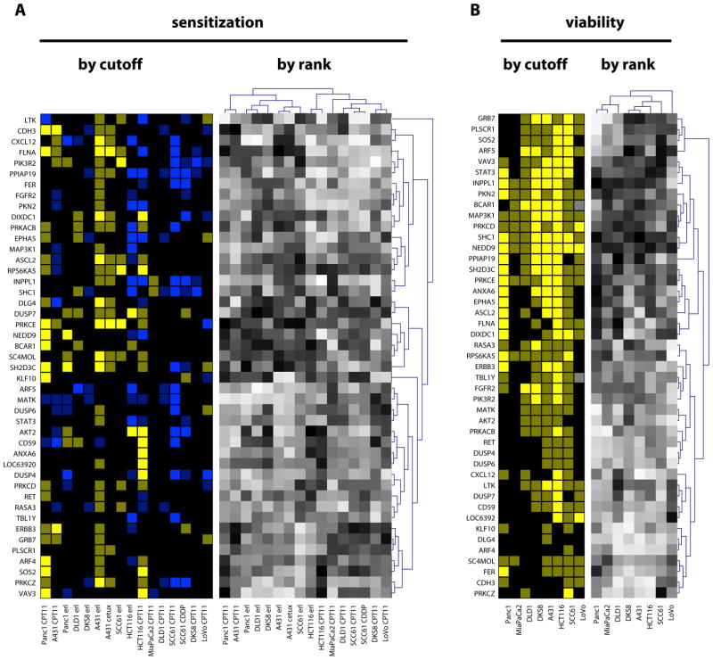
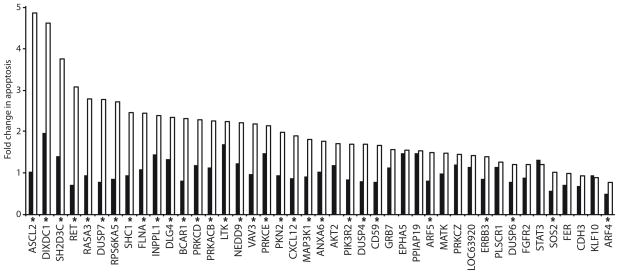
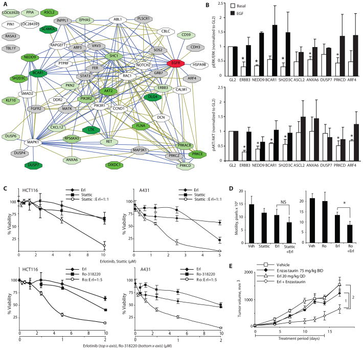
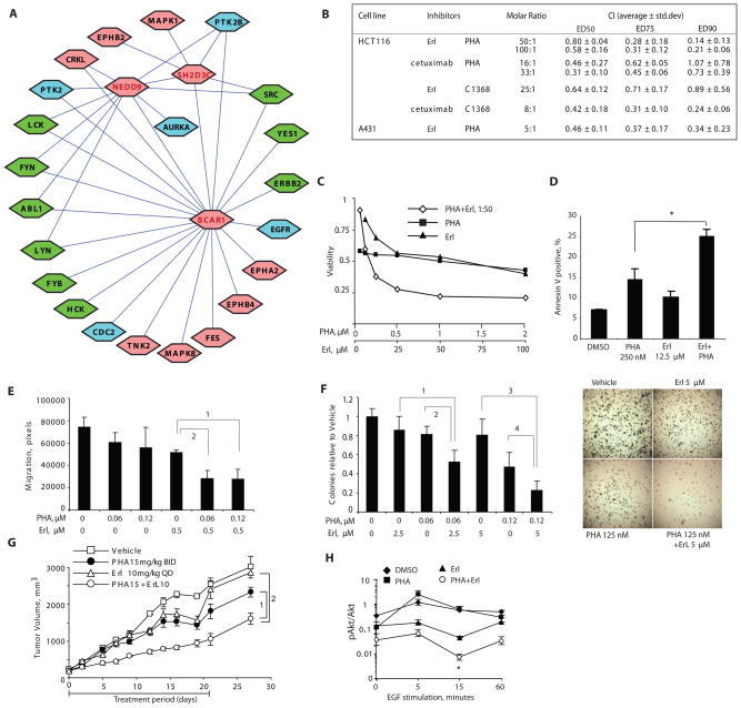
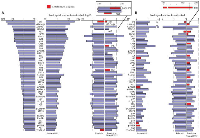
Intrinsic and acquired cellular resistance factors limit the efficacy of most targeted cancer therapeutics. Synthetic lethal screens in lower eukaryotes suggest that networks of genes closely linked to therapeutic targets would be enriched for determinants of drug resistance. We developed a protein network centered on the epidermal growth factor receptor (EGFR), which is a validated cancer therapeutic target, and used small interfering RNA screening to comparatively probe this network for proteins that regulate the effectiveness of both EGFR-targeted agents and nonspecific cytotoxic agents. We identified subnetworks of proteins influencing resistance, with putative resistance determinants enriched among proteins that interacted with proteins at the core of the network. We found that clinically relevant drugs targeting proteins connected in the EGFR network, such as protein kinase C or Aurora kinase A, or the transcriptional regulator signal transducer and activator of transcription 3 (STAT3), synergized with EGFR antagonists to reduce cell viability and tumor size, suggesting the potential for a direct path to clinical exploitation. Such a focused approach can potentially improve the coherent design of combination cancer therapies.


Figures

Fig. 1
Fig. 1. Design and screening of a targeted library
(A) Genes targeted in the library were identified from a preliminary large set (open circles) of genes engaged in protein-protein interactions (PPIs) with one of the seeds, or interacting with one of the direct interactors; collected from 5 curated pathway resources for the EGFR signaling network (Pathway maps); shown by microarray experiments to be rapidly transcriptionally responsive to stimulation or inhibition of EGFR; or human orthologs of genes known to genetically interact with EGFR in Drosophila (Drosophila genetics). Genes from this larger set were prioritized by a series of collection criteria aimed at finding genes connected to EGFR by more than one criterion (detailed in table S1); final numbers of genes added to the library from each group are shown in filled circles. At the intersection of the circles is the number of genes added to the library, because they belong to at least two sets. For the PPI, pathway map, and microarray sets, additional 77 genes were identified as closely related in sequence and potentially paralogous in function (dashed line). (B) Distribution of hits as a factor of overall viability reduction with the siRNA. siRNAs in library are listed in order of intrinsic impact on viability of A431 cells treated with DMSO (gray line). Blue triangles, sensitization index (SI) for primary hits with erlotinib; red triangles, SI for validated hits with erlotinib; green squares, SI for primary hits with CPT11. (C) Degree of overlap between primary hits obtained for erlotinib, panitumumab, and CPT11. (D) Network of validated (red circles) hits sensitizing to EGFR-targeting agents, in the context of the full library. Lines (edges) represent connections based on direct protein-protein interactions or genetic interactions in Drosophila. Hits and genes from the starting set that were not connected in the network are shown below the network.
Fig. 2
Fig. 2. Network properties of hits
(A) Hits by source of input in library. MA, microarray indicates transcriptionally responsive to EGFR; DG, Drosophila genetics; PPI1, direct protein interactions with seeds; PM, pathway maps; PPI2, direct protein interactions with a protein within the PPI1 group, or found in a complex with seed proteins; 3S, any 3 sources combined. (B) Topological analysis of erlotinib hit network identified in A431 cells. Data shown represents difference between properties of the set of 61 validated hits and the average for 20 randomly generated sets of 61 genes from the library. Measures of degree, topological coefficient (Topological Coeff/100), stress (Stress/1000), and neighborhood connectivity (Neighborhood Conn/3) show significant enrichment for hits validated with erlotinib, with the error bars for the random set data reflecting a 99% confidence interval. (C) Enriched GO classifications (for each category shown, p<0.01). among hits. Enrichment is significant for proteins annotated as involved in phosphate metabolism (kinases and phosphatases) and in multiple signal transduction-related categories. (D) Percentage of hits versus library proteins having a recognized ortholog in S. cerevisiae, C. elegans or D. melanogaster. X axis, the number of species (among listed) having a recognized ortholog.
Fig. 3
Fig. 3. Sensitization profile of hits
(A) Left, SI values for erlotinib and CPT11, calculated as (test siRNA)/(GL2) for cells grown in drug, divided by (test siRNA/GL2) for cells grown in DMSO: dim yellow, SI≤0.85; bright yellow, SI≤0.7; dim blue, SI≥1.15; bright blue, SI≥1.3. Right, gradient of relative ranking of the efficacy of hits to sensitize cells to drugs indicated: black, most sensitizing; white, least sensitizing or in some cases antagonizing (compare to left panels). For rank order analysis, cluster analysis was performed to identify genes with similar profiles of sensitization (dendrogram, Y axis), and also to cluster cell lines by similarity of response (dendrogram, X axis): these clustered patterns are used to organize the display of genes selected by threshold. Two cell lines, MIA-Paca2 and LoVo, yielded no reproducible hits sensitizing to erlotinib; BCAR1 and TBL1Y were not tested in LoVo. Note, not all hits initially obtained and validated in A431 score as positive by rigorous statistical criteria with A431/erlotinib in this experiment because of the intrinsic false negative rate of the assay. (B) Left, siRNA-induced viability reduction below 0.85 (dim yellow), or 0.7 (bright yellow) that of control siRNA-treated cells, calculated based on the formula (test siRNA)/(GL2 control siRNA) for cells grown in DMSO. Right, relative ranking of hit efficacy in reducing cell line viability.
Figure 4
Figure 4. siRNA hits enhance apoptosis in the presence of EGFR inhibitor
Apoptosis was determined by annexin-V labeling and normalized to negative control siRNA. Composite results from two independent experiments are shown as odds ratio columns; black, DMSO-treated; white, erlotinib-treated. Asterisks indicate statistically significant (FDR ≤ 0.05) erlotinib-gene interactions from two independent experiments.
Fig. 5
Fig. 5. Functional classification of hits
(A) Network of validated genes that sensitize cells to EGFR-targeting agents. Blue lines represent connections based on direct PPIs or genetic interactions in Drosophila; green lines represent connections generated by pathway maps and text mining. Green shadowing on boxes indicates genes that are in the top quartile by rank order of those strongly sensitizing at least 1 (lightest green) to at least 5 (darkest green) cancer cell lines to erlotinib. Gray boxes, genes that were never in the top quartile; white boxes, validated genes not tested for sensitization. (B) Analysis of ERK (top) and AKT (bottom) status in cell lysates from A431 cells following siRNA transfection, under basal medium conditions and following EGF stimulation. Average results of three independent Western blots are shown as ratios of amounts of phosphorylated and total proteins. Results were normalized to corresponding ratios in GL2 siRNA-transfected control cells; error bars are standard deviations. Asterisks indicate statistically significant difference with negative control; t-test p<0.05. (C) Viability curves for erlotinib and Stattic used as single agents, or combined at 1:1 molar ratio in A431 (left) and HCT116 (right) cells, or for erlotinib and Ro-318220 used as single agents, or combined at 1:5 molar ratio in A431 (left) and HCT116 (right) cells. (D) Motility was measured by wound healing assay in A431 cells cells treated with 0.5 μM erlotinib alone or in combination with 0.5 μM Stattic (left) or 0.25 μM Ro-318220 (right), and assessed over 18 hours. NS, not significant. *, FDR=3.57*10−5. (E) A431 xenografts implanted in SCID mice were treated with drugs as indicated when palpable tumors appeared (day zero) Drug treatment was administered from days 0–14, and tumor measurement continued until day 18. The generalized estimating equations approach (with an autoregressive correlation structure) was used to model tumor growth. A linear time-effect was included in the model for the logarithm of tumor volume and interacted with the treatments in each comparison. P-values are 1, 0.042 and 2, 0.032, n=5 mice per group.
Fig. 6
Fig. 6. Synergy between inhibitors of AURKA and inhibitors of EGFR
(A) Kinases directly associated with the BCAR1-NEDD9-SH2D3C cluster. Kinases for which no clinical small molecule inhibitor is available are indicated in pink; kinases for which small molecule inhibitors are available are indicated in blue or in green, if inhibitor has previously been tested for synergy with EGFR-targeting agents. (B) Inhibitors of EGFR and inhibitors of AURKA synergize to reduce viability of cancer cells in vitro. Summary results of drug interactions calculated as Chou-Talalay combination index (CI) based on Cell Titer blue viability determinations. CI values <1 indicate synergy, and <0.5 strong synergy, between the two agents in producing cytotoxic effect.. (C) Dose-dependent inhibition of HCT116 cell viability by combination of PHA-680632 and erlotinib. (D) Combination of PHA-680632 and erlotinib treatment increases apoptosis in HCT116 cells at 72 hours; *, t-test p=0.001. (E) Cell motility was measured by wound healing assay in cells treated with drugs at concentrations indicated, and assessed over 18 hours. FDR is <10−5 for 1 and 2. (F) Left, relative soft agar colony formation of cells grown for 2 to 3 weeks in drugs at the concentrations indicated. FDR is equal to 1, 0.0003; 2, 0.0006; 3, 0.0003; and 4, 0.004. Right, results from typical experiment. (G) Tumor xenografts were implanted in SCID mice treated with drugs as indicated. The generalized estimating equations approach (with an autoregressive correlation structure) was used to model tumor growth. A linear time-effect was included in the model for the logarithm of tumor volume and interacted with the treatments in each comparison. P-values are 1: 0.005; 2: 0.008. (H) Quantitation of 3 independent Western analyses of protein lysates of cells treated with erlotinib and PHA-680632 for 3 hrs followed by EGF stimulation. Error bars represent standard error of the mean (SEM) of three independent experiments; *, t-test comparing erlotinib to erlotinib + PHA-680632 yielded P = 0.013.
Fig. 7
Fig. 7. Dual inhibition of Aurora-A and EGFR suppresses activation of multiple signaling nodes
Ranked fold increase in phosphorylation signal of 46 proteins in A431 cells stimulated with EGF (15 ng/mL, 15 min) (A) or grown in 1% serum media (B) and treated with indicated drugs. Stimulation in cells exposed to EGF in the absence of drugs is shown in the far left of panel A. In panel A, the proteins are listed in the same order in the right and left sides, with all proteins labeled on the left and those that are substantially reduced by drug treatment labeled on the right. In panel A, cells were exposed to 0.5 μM erlotinib or 1 μM PHA-680632, or both. In panel B, cells were exposed to 1 μM erlotinib or 0.5 μM PHA-680632, or 0.5 μM erlotinib plus 1 μM PHA-680632. Inset, graphs illustrate magnified scale of indicated phosphoproteins. Proteins showed in red have consistent decrease of >2-fold in signal intensity in independent biological replicates, as indicated.

References

    1. Pao W, Wang TY, Riely GJ, Miller VA, Pan Q, Ladanyi M, Zakowski MF, Heelan RT, Kris MG, Varmus HE. KRAS mutations and primary resistance of lung adenocarcinomas to gefitinib or erlotinib. PLoS medicine. 2005 Jan;2:e17. - PMC - PubMed
    1. Lievre A, Bachet JB, Le Corre D, Boige V, Landi B, Emile JF, Cote JF, Tomasic G, Penna C, Ducreux M, Rougier P, Penault-Llorca F, Laurent-Puig P. KRAS mutation status is predictive of response to cetuximab therapy in colorectal cancer. Cancer Res. 2006 Apr 15;66:3992. - PubMed
    1. Friedman A, Perrimon N. Genetic screening for signal transduction in the era of network biology. Cell. 2007 Jan 26;128:225. - PubMed
    1. Barabasi AL, Albert R. Emergence of scaling in random networks. Science. 1999 Oct 15;286:509. - PubMed
    1. Hartwell LH, Hopfield JJ, Leibler S, Murray AW. From molecular to modular cell biology. Nature. 1999 Dec 2;402:C47. - PubMed

Publication types

MeSH terms