. 2010 Nov 26;285(48):37370-6.
doi: 10.1074/jbc.M110.170084. Epub 2010 Sep 21.

Ca2+ overload and sarcoplasmic reticulum instability in tric-a null skeletal muscle

Affiliations

Ca2+ overload and sarcoplasmic reticulum instability in tric-a null skeletal muscle

Xiaoli Zhao et al. J Biol Chem. .

Abstract

The sarcoplasmic reticulum (SR) of skeletal muscle contains K(+), Cl(-), and H(+) channels may facilitate charge neutralization during Ca(2+) release. Our recent studies have identified trimeric intracellular cation (TRIC) channels on SR as an essential counter-ion permeability pathway associated with rapid Ca(2+) release from intracellular stores. Skeletal muscle contains TRIC-A and TRIC-B isoforms as predominant and minor components, respectively. Here we test the physiological function of TRIC-A in skeletal muscle. Biochemical assay revealed abundant expression of TRIC-A relative to the skeletal muscle ryanodine receptor with a molar ratio of TRIC-A/ryanodine receptor ∼5:1. Electron microscopy with the tric-a(-/-) skeletal muscle showed Ca(2+) overload inside the SR with frequent formation of Ca(2+) deposits compared with the wild type muscle. This elevated SR Ca(2+) pool in the tric-a(-/-) muscle could be released by caffeine, whereas the elemental Ca(2+) release events, e.g. osmotic stress-induced Ca(2+) spark activities, were significantly reduced likely reflecting compromised counter-ion movement across the SR. Ex vivo physiological test identified the appearance of "alternan" behavior with isolated tric-a(-/-) skeletal muscle, i.e. transient and drastic increase in contractile force appeared within the decreasing force profile during repetitive fatigue stimulation. Inhibition of SR/endoplasmic reticulum Ca(2+ ATPase) function could lead to aggravation of the stress-induced alternans in the tric-a(-/-) muscle. Our data suggests that absence of TRIC-A may lead to Ca(2+) overload in SR, which in combination with the reduced counter-ion movement may lead to instability of Ca(2+) movement across the SR membrane. The observed alternan behavior with the tric-a(-/-) muscle may reflect a skeletal muscle version of store overload-induced Ca(2+) release that has been reported in the cardiac muscle under stress conditions.


Figures

FIGURE 1.
FIGURE 1.
Abundant expression of TRIC protein in skeletal muscles. A, representative Coomassie Brilliant Blue staining showed the relative expression levels of RyR (552 kDa) and SERCA protein (100 kDa) (left panel), as well as CSQ (60 kDa) and TRIC-A protein (33 kDa) (middle panel) in isolated SR microsome from rabbit skeletal muscle, as compared with the BSA standard. Right panel was a Western blot of the isolated microsome from rabbit hindlimb skeletal muscle using rabbit polyclonal anti-TRIC-A antibody. B, representative Western blot showing the differential expression patterns of TRIC-A and TRIC-B in hindlimb, soleus (SOL), and EDL from the wild type mice. C, summarized data of the relative expression levels of TRIC-A and TRIC-B in hindlimb (n = 8), soleus (n = 8), and EDL (n = 8) from wild type skeletal muscle. The expression levels of TRIC-A and TRIC-B in hindlimb were set as 100%, **, p < 0.01.
FIGURE 2.
FIGURE 2.
Detection of large-size vacuoles containing electron-dense Ca2+ deposit in tric-a−/− muscles. A, toluidine blue-stained light microscopic cross-section image of a tric-a−/− muscle bundle at low magnification, arrows designate myofibrils containing large-sized vacuoles. B, enlarged image of a single tric-a−/− myofibril containing large-sized vacuoles. C, transmission EM image stained by oxidized iron showed the longitudinal thick section of tric-a−/− FDB at high magnification. Mitochondria (Mit) was labeled in the image and arrows designate electron dense Ca2+ deposits in the large-sized vacuoles (>0.5 μm in diameter), preferentially formed in the terminal SR region. D, enlarged image showing a Ca2+-overloaded swollen SR. E, transmission EM image stained by oxidized iron showed the longitudinal thick section of wild type FDB at high magnification.
FIGURE 3.
FIGURE 3.
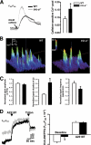
Altered SR Ca2+ release properties and membrane potential in tric-a mutant muscles. A, representative trace of intracellular Ca2+ transient induced by 20 mm caffeine (arrow) in FDB from wild type (gray) and tric-a−/− (black) mice (left panel). Fura-2 fluorescence was recorded at excitation wavelengths of 350 (F350) and 380 nm (F380). The peak caffeine response at F350/F380 in wild type (open bar) and tric-a−/− muscle (filled bar) is summarized in the right panel, n = 10. B, representative three-dimensional images of Ca2+ spark in wild type and tric-a−/− FDB fibers. C, normalized amplitude (ΔF/F0), duration of single spark (ms), and frequency per min of osmotic-induced Ca2+ spark in FDB of the wild type (open) and tric-a−/− mice (filled) were summarized here, n = 13–24. The value of wild type fibers were set as 1. D, representative trace of SR membrane potential measurement by di-8-ANEPPQ dye at F510/F450 (left panel) in SR vesicle isolated from wild type (gray) and tric-a−/− (black) mice, and arrows designate when A23187 (Ca2+ ionophore) and Triton were added (left panel). Right panel shows summarization of this data, F510/F450 after addition of Triton was set as 0, n = 6. All values were mean ± S.E., *, p < 0.05.
FIGURE 4.
FIGURE 4.
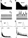
Mechanical alternans in TRIC-A absent muscles induced by fatigue stimulation. A, isolated muscle bundles from wild type mice show monotonic decrease in contractile force, when subjected to fatiguing stimulations by the 50% Tmax frequency. B, isolated muscle bundles from the tric-a−/− mice show irregular patterns contractile force, when subjected to similar fatiguing stimulations as in A. Enlarged individual contractile forces shown in the lower panels reveal the appearance of alternans in the tric-a−/− muscle. C, EDL muscles from the wild type mice were treated with 20 μm CPA for 5 min, and tetanic contractile force measurements reveal the appearance of the characteristic slow relaxing phase (top panel). In the presence of CPA, fatiguing stimulation leads to the appearance of mild alternans in wild type muscle (bottom panel). The trace is representative of 4 of 10 muscle preparations. D, representative traces for the tric-a−/− muscle as shown using identical experimental conditions as in C. Clearly, CPA treatment leads to aggravation of alternan events in the tric-a−/− muscle. Trace shown at the bottom panel of D is representative of 8 of 10 muscle preparations tested.

References

    1. Ríos E., Ma J. J., González A. (1991) J. Muscle Res. Cell Motil. 12, 127–135 - PubMed
    1. Melzer W., Herrmann-Frank A., Lüttgau H. C. (1995) Biochim. Biophys. Acta 1241, 59–116 - PubMed
    1. Fill M., Copello J. A. (2002) Physiol. Rev. 82, 893–922 - PubMed
    1. Takeshima H., Komazaki S., Nishi M., Iino M., Kangawa K. (2000) Mol. Cell 6, 11–22 - PubMed
    1. Hirata Y., Brotto M., Weisleder N., Chu Y., Lin P., Zhao X., Thornton A., Komazaki S., Takeshima H., Ma J., Pan Z. (2006) Biophys. J. 90, 4418–4427 - PMC - PubMed

Publication types