. 2010 Nov 26;285(48):37811-22.
doi: 10.1074/jbc.M110.127498. Epub 2010 Sep 21.

Identification of the Drosophila ortholog of HSPB8: implication of HSPB8 loss of function in protein folding diseases

Affiliations

Identification of the Drosophila ortholog of HSPB8: implication of HSPB8 loss of function in protein folding diseases

Serena Carra et al. J Biol Chem. .

Abstract

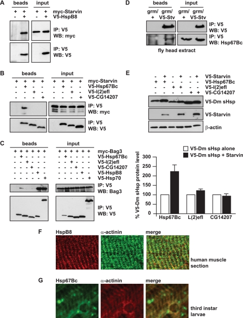
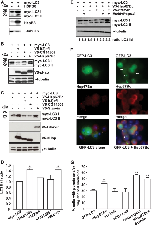
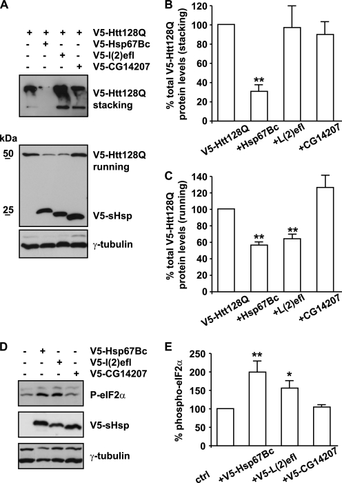
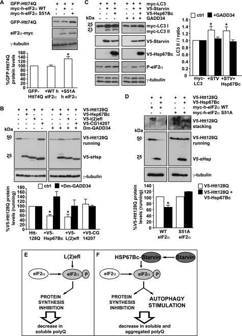
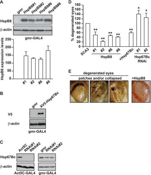
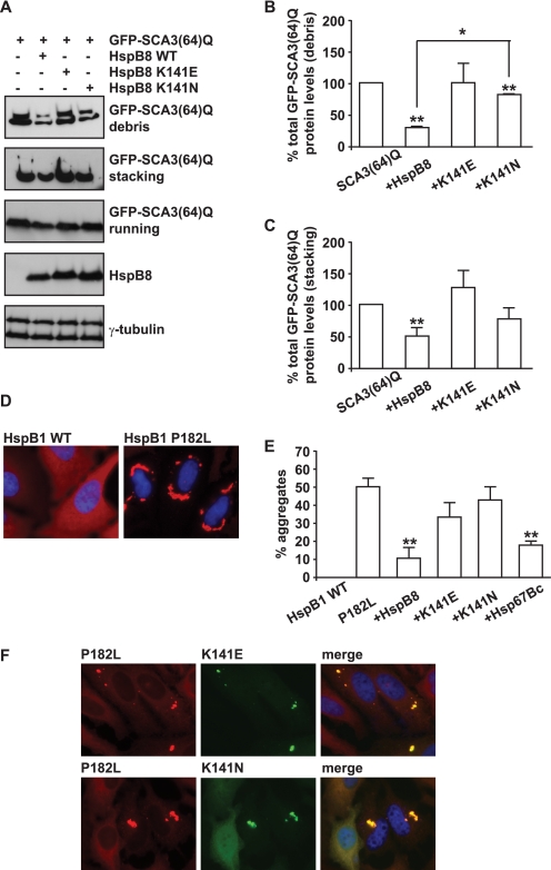
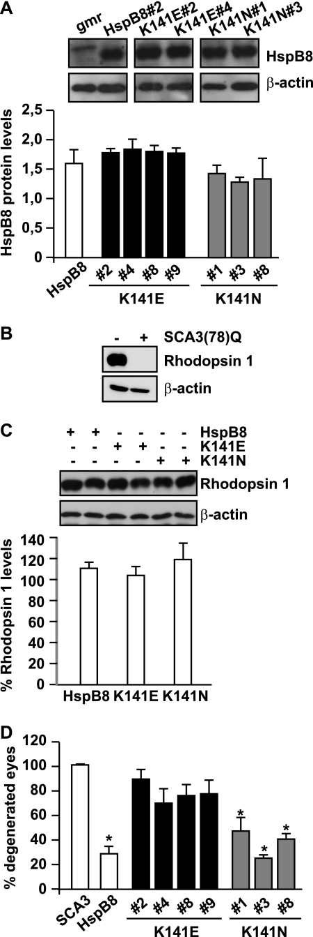
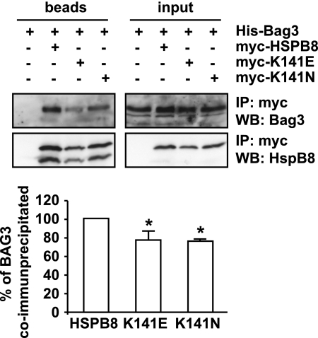
Protein aggregation is a hallmark of many neuronal disorders, including the polyglutamine disorder spinocerebellar ataxia 3 and peripheral neuropathies associated with the K141E and K141N mutations in the small heat shock protein HSPB8. In cells, HSPB8 cooperates with BAG3 to stimulate autophagy in an eIF2α-dependent manner and facilitates the clearance of aggregate-prone proteins (Carra, S., Seguin, S. J., Lambert, H., and Landry, J. (2008) J. Biol. Chem. 283, 1437-1444; Carra, S., Brunsting, J. F., Lambert, H., Landry, J., and Kampinga, H. H. (2009) J. Biol. Chem. 284, 5523-5532). Here, we first identified Drosophila melanogaster HSP67Bc (Dm-HSP67Bc) as the closest functional ortholog of human HSPB8 and demonstrated that, like human HSPB8, Dm-HSP67Bc induces autophagy via the eIF2α pathway. In vitro, both Dm-HSP67Bc and human HSPB8 protected against mutated ataxin-3-mediated toxicity and decreased the aggregation of a mutated form of HSPB1 (P182L-HSPB1) associated with peripheral neuropathy. Up-regulation of both Dm-HSP67Bc and human HSPB8 protected and down-regulation of endogenous Dm-HSP67Bc significantly worsened SCA3-mediated eye degeneration in flies. The K141E and K141N mutated forms of human HSPB8 that are associated with peripheral neuropathy were significantly less efficient than wild-type HSPB8 in decreasing the aggregation of both mutated ataxin 3 and P182L-HSPB1. Our current data further support the link between the HSPB8-BAG3 complex, autophagy, and folding diseases and demonstrate that impairment or loss of function of HSPB8 might accelerate the progression and/or severity of folding diseases.


Figures

FIGURE 1.
FIGURE 1.
D. melanogaster HSP67Bc is a functional ortholog of human HSPB8. A and B, human HSPB8 and Dm-HSP67Bc co-immunoprecipitate with Dm-Starvin. HEK-293T cells were transfected with vectors encoding for D. melanogaster Myc-Starvin alone or together with either V5-HSPB8, V5-HSP67Bc, V5-L(2)efl, or V5-CG14207. 24 h post-transfection, the cell lysates were subjected to immunoprecipitation (IP) with an antibody against the V5 tag, and the immunoprecipitated complexes were analyzed by Western blotting (WB) using V5- and Myc-specific antibodies. Among the D. melanogaster sHSPs analyzed, HSP67Bc interacts with Dm-Starvin (B), similarly to human HSPB8 (A). C, like HSPB8, Dm-HSP67Bc also binds to BAG3, the human functional ortholog of Dm-Starvin. HEK293 cells were transfected with vectors encoding for human Myc-BAG3 alone or together with either V5-HSP67Bc, V5-L(2)efl, V5-CG14207, or V5-HSPB8 and V5-HSP70, both used as positive controls and subjected, 24 h post-transfection, to immunoprecipitation with a V5-specific antibody. D, endogenous Hsp67Bc interacts with Starvin in vivo in fly head extracts. V5-starvin was expressed in flies under the control of the grm-GAL4 driver. Immunoprecipitation with a specific V5 antibody was carried out using fly head protein extracts from control flies (grm/+) and flies expressing V5-Starvin (gmr/V5-Stv). Interaction of endogenous Hsp67Bc with V5-Starvin was investigated by Western blotting using a specific rabbit polyclonal Hsp67Bc antibody. E, total levels of HSP67Bc are increased when it is co-expressed with Starvin. Drosophila Schneider S2 cells were transfected with vectors encoding for V5-HSP67Bc, V5-L(2)efl, and V5-CG14207 alone or in combination with V5-Starvin. The protein expression levels were analyzed by Western blotting 48 h post-transfection (average values ± S.E. (error bars) of n = 3–4 independent samples). F, human muscle tissue section showing that endogenous HSPB8 colocalizes with α-actinin at the Z band. G, endogenous Dm-HSP67Bc colocalizes with α-actinin at the Z band in third instar larvae muscles.
FIGURE 2.
FIGURE 2.
HSP67Bc and Starvin induce LC3 lipidation both in mammalian and Drosophila Schneider S2 cells. A, HSPB8 overexpression leads to an increase in the total LC3 I and LC3 II levels. HEK293T cells were transfected with vectors encoding for Myc-LC3 alone or together with human HSPB8. 44 h post-transfection, the cell lysates were analyzed by Western blotting using Myc- and HSPB8-specific antibodies. B–D, HEK293T cells were transfected with vectors encoding for Myc-LC3 alone or together with either V5-HSP67Bc, V5-L(2)efl, V5-CG14207, or V5-Starvin. 44 h post-transfection, the cell lysates were analyzed by Western blotting using Myc- and V5-specific antibodies. Endogenous levels of γ-tubulin were measured as loading control. B and C, HSP67Bc and Starvin, but not L(2)efl or CG14207, induce LC3 lipidation. D, quantification of the effect of Hsp67Bc, L(2)efl, CG14207, and Starvin on the LC3 II/LC I ratio. *, p < 0.05; average values ± S.E. (error bars) of n = 3–4 independent samples. E, overexpression of HSP67Bc alone or together with Starvin induces the turnover of LC3. Where indicated (+), cells were treated for 6 h with the lysosomal inhibitors pepstatin A and E64d prior to extraction. Quantification of the effect of the lysosomal inhibitors on the LC3 II/LC3 I ratio (normalized against γ-tubulin) in cells transfected for 36 h is shown. F and G, overexpression of HSP67Bc alone or together with Starvin significantly increases the number of S2 cells containing GFP-LC3-positive punctae or ring-shaped vacuole-like structures. Drosophila Schneider S2 cells were transfected with vectors encoding for GFP-LC3 alone or together with either V5-HSP67Bc, V5-L(2)efl, V5-CG14207, or V5-Starvin or with both V5-HSP67Bc and V5-Starvin. 24 h post-transfection, cells were either left untreated or treated with rapamycin (5 μm) for 180 min and then fixed with formaldehyde at room temperature for 10 min. F, low magnification image showing the induction of GFP-LC3-positive autophagic vacuoles (white arrows) by HSP67Bc. G, the number of cells containing GFP-LC3-positive punctae and/or vacuole-like structures was counted. *, p < 0.05; **, p < 0.001; average values ± S.E. of n = 4–11 independent samples.
FIGURE 3.
FIGURE 3.
HSP67Bc inhibits the accumulation of both soluble and high molecular weight insoluble mutated huntingtin in Drosophila Schneider S2 cells. A, Drosophila Schneider S2 cells were transfected with vectors encoding for mutated huntingtin with a 128-glutamine repeat (V5-Htt128Q) alone or together with either V5-HSP67Bc, V5-L(2)efl, or V5-CG14207, and total proteins were extracted 48 h post-transfection. Quantification of the effect of the chaperones on the accumulation of the high molecular weight insoluble forms retained in the stacking gel (B) and the soluble monomeric V5-Htt128Q (C) is shown. **, p < 0.001; average values ± S.E. (error bars) of n = 4–6 independent samples. D, HEK-293 cells were transfected with vectors encoding for either mRFP, V5-HSP67Bc, V5-L(2)efl, or V5-CG14207; total proteins were extracted 24 h post-transfection, and levels of phospho-eIF2α were measured by Western blotting. E, quantification of the effect of the chaperones on the levels of phospho-eIF2α. **, p < 0.001; *, p < 0.05; average values ± S.E. of n = 3 independent samples.
FIGURE 4.
FIGURE 4.
Similarly to human HSPB8, the ability of HSP67Bc to decrease mutated huntingtin accumulation/aggregation and to induce autophagy is eIF2α-dependent. A, impairment of the eIF2α pathway increases mutated polyglutamine protein levels in Drosophila Schneider S2 cells. Drosophila Schneider S2 cells were transfected with vectors encoding for GFP-Htt74Q and either Myc-tagged human wild-type eIF2α or Myc-tagged non-phosphorylatable human eIF2α S51A. 48 h post-transfection, the total levels of GFP-Htt74Q were quantified (*, p < 0.05; average values ± S.E. (error bars) of n = 3 independent samples). B, HSP67Bc and L(2)efl decrease mutated huntingtin soluble levels via the eIF2α pathway. Drosophila Schneider S2 cells were transfected with vectors encoding for V5-Htt128Q alone or together with either V5-HSP67Bc, V5-L(2)efl, or V5-CG14207. Where indicated (+), a vector encoding for D. melanogaster GADD34 (Dm-GADD34) was also co-transfected. Total proteins were extracted 48 h later, and V5-Htt128Q soluble (running) levels were analyzed by Western blotting. *, p < 0.05; average values ± S.E. of n = 3 independent samples. C, the induction of LC3 lipidation by HSP67Bc and Starvin is dependent on the phosphorylation of eIF2α. HEK293T cells were transfected with vectors encoding for Myc-LC3 alone or together with V5-Starvin or V5-Starvin and V5-HSP67Bc. Where indicated (+), cells were cotransfected with a vector encoding for the C terminus of GADD34, which inhibits the phosphorylation of endogenous eIF2α. Cell lysates were prepared 44 h post-transfection and analyzed by Western blotting. *, p < 0.05; average values ± S.E. of n = 3–4 independent samples. D, S51A eIF2α completely abrogated the ability of HSP67Bc to decrease V5-Htt128Q soluble (running) and insoluble (stacking) levels. Drosophila Schneider S2 cells were transfected with vectors for V5-Htt128Q alone or together with V5-HSP67Bc and either human wild-type Myc-eIF2α or the non-phosphorylatable mutant S51A. Total proteins were extracted 48 h later. Quantification of the soluble (running) protein levels of V5-Htt128Q normalized against endogenous γ-tubulin is shown (*, p < 0.01; average values ± S.E. of n = 3 independent samples). E and F, schematic model showing the putative mechanism of action of L(2)efl and HSP67Bc. E, overexpression of L(2)efl leads to the phosphorylation of eIF2α, thereby causing a translational shutdown. This will result in a decrease of the levels of soluble poly(Q) proteins, without affecting significantly the rate of their aggregation. F, overexpression of HSP67Bc, which interacts with Starvin, also leads to the phosphorylation of eIF2α, which, however, causes both translational shutdown and stimulation of autophagy. As a consequence, HSP67Bc-Starvin leads to a decrease of the levels of soluble poly(Q) proteins, but it also facilitates the clearance of aggregated poly(Q) proteins by stimulating autophagy.
FIGURE 5.
FIGURE 5.
Overexpression of human HSPB8 or Dm-HSP67Bc protects against mutated SCA3-induced eye degeneration in vivo. grm-GAL4-driven expression levels of human HSPB8 (transgenic lines 1, 2, 6, and 8; A) and of V5-HSP67Bc (B). The expression levels of the different transgenic lines expressing human HSPB8 were not significantly different. GMR, gmr-GAL4/+; HSPB8#1, gmr-GAL4/UAS-HSPB8#1, etc.; V5-HSP67Bc, gmr-GAL4/+; UAS-V5-HSP67Bc/+. C, Act5C-GAL4-driven and gmr-GAL4-driven knockdown of Dm-HSP67Bc with two independent UAS RNAi lines (VDRC transformant lines ID 26416 and 26417) grown at 25 °C showing decreased expression of the endogenous protein. Protein levels were measured using protein extracts from 1–2-day-old fly heads. Left, Act5C, Act5C-GAL4/+; RNAi#1, Act5C-GAL4/+; UAS-CG4190 RNAi#1/+; RNAi#2, Act5C-GAL4/+; UAS-CG4190 RNAi#2/+. Right, gmr, gmr-GAL4/+; RNAi#1, gmr-GAL4/+; UAS-CG4190 RNAi#1/+; RNAi#2, gmr-GAL4/+; UAS-CG4190 RNAi#2/+. D, quantification of eye degeneration in flies overexpressing either SCA3(78Q) alone (or in combination with HSPB8 or HSP67Bc). Co-expression of human HSPB8 or V5-HSP67Bc significantly decreases the percentage of flies with degenerated eyes (dark patches and/or collapsed eyes) as compared with control flies, whereas knockdown of endogenous HSP67Bc enhances eye degeneration. SCA3, gmr-GAL4-UAS-SCA3(78Q)/+; HSPB8#1, gmr-GAL4-UAS-SCA3(78Q)/UAS-HSPB8#1, etc.; HSP67Bc, gmr-GAL4-UAS-SCA3(78Q)/+; UAS-V5-HSP67Bc/+; HSP67Bc RNAi#1, gmr-GAL4-UAS-SCA3(78Q)/+; UAS-CG4190 RNAi#1/+; HSP67Bc; RNAi#2, gmr-GAL4-UAS-SCA3(78Q)/+; UAS-CG4190 RNAi#2/+. Total number of eyes scored was 200–400; **, p < 0.001; *, p < 0.05; average values ± S.E. (error bars) of n = 3 independent experiments. E, representative picture showing SCA3(78Q) flies with degenerated eyes and the partial rescue obtained by overexpression of human HSPB8.
FIGURE 6.
FIGURE 6.
In cells, the K141E and K141N mutations show impaired ability to decrease the aggregation of the two misfolded substrates SCA3(64)Q and P182L-HSPB1 as compared with wild-type HSPB8. A–C, K141E and K141N are less efficient than wild-type HSPB8 in decreasing mutated ataxin 3 aggregation and mediated toxicity. HEK293T cells were transfected with vectors encoding for SCA3(64)Q alone or together with either wild-type HSPB8, K141E, or K141N. 48 h post-transfection, the medium (debris) and the cells were collected, and total proteins from both fractions were extracted. SCA3(64)Q soluble (running) and insoluble (stacking) levels were analyzed. B and C, total levels of GFP-SCA3(64)Q were quantified (*, p < 0.05; average values ± S.E. (error bars) of n = 3 independent samples). D and E, K141E and K141N are less efficient than wild-type HSPB8 in decreasing mutated P182L-HSPB1 aggregation. HeLa cells were transfected with vectors encoding for wild-type or P182L-HSPB1 or with vectors encoding for P182L-HSPB1 alone or in combination with wild-type Myc-HSPB8, Myc-K141E, Myc-K141N, or wild-type Myc-HSP67Bc. 72 h post-transfection, cells were fixed with 3.7% formaldehyde and processed for immunofluorescence with specific antibodies. E, the number of cells showing HSPB1 aggregates was counted (*, p < 0.05; average values ± S.E. of n = 3–5 independent samples; more than 100 cells/sample were counted). F, mutated K141E and K141N colocalize with aggregated P182L-HspB1.
FIGURE 7.
FIGURE 7.
The K141E mutation significantly affects HSPB8 protective effect on SCA3-induced eye degeneration. A, quantification of gmr-GAL4-driven HSPB8 protein expression in 1–3-day-old fly head extracts (mean ± S.E. (error bars); n = 3–7). Expression of human wild-type HSPB8 (transgenic line 2), used as control, mutated HSPB8 K141E (transgenic lines 2, 4, 8, and 9) and K141N (transgenic lines 1, 3, and 8). gmr, gmr-GAL4/+; HSPB8#2, gmr-GAL4/UAS-HSPB8#2; K141E#2, gmr-GAL4/UAS-HSPB8-K141E#2, etc.; K141N#1, gmr-GAL4/UAS-HSPB8-K141N#1, etc. B, rhodopsin 1 levels are decreased below detectable levels in SCA3(78)Q-flies and can be used as a marker for eye degeneration. Protein extracts from 1–2-day-old fly heads, with corresponding genotypes: −, CyO/+; +, gmr-GAL4-UAS-SCA3 (78Q)/+. C, overexpression of mutated HSPB8 alone does not significantly affect rhodopsin 1 levels. Protein extracts were prepared from 20 days old fly heads. Western blotting of two representative samples per group and quantification of rhodopsin 1 levels normalized against β-actin show no significant decrease upon overexpression of mutated HSPB8, as compared with wild-type HSPB8 (rhodospin 1 levels from 3–4 independent lines expressing either wild-type HSPB8 or K141E or K141N were quantified, and measures were pooled; mean ± S.E.; n = 7–13). Genotypes were as described in the legend to Fig. 6A. D, mutations of HSPB8 decrease its ability to protect against mutated SCA3-mediated eye degeneration. Shown is a quantification of eye degeneration in flies overexpressing either SCA3(78Q) alone or in combination with wild-type or mutated, K141E or K141N, HSPB8; total number of eyes scored: 200–300; *, p < 0.05 as compared with SCA3; average values ± S.E. of n = 3 independent experiments. SCA3, gmr-GAL4-UAS-SCA3(78Q)/+; HSPB8, gmr-GAL4-UAS-SCA3(78Q)/UAS-HSPB8#2; K141E#2, gmr-GAL4-UAS-SCA3(78Q)/UAS-HSPB8-K141E#2, etc; K141N#1, gmr-GAL4-UAS-SCA3(78Q)/UAS-HSPB8-K141N#1, etc.
FIGURE 8.
FIGURE 8.
The K141E and K141N mutated forms bind less strongly to BAG3 than wild-type HSPB8. HEK-293T cells were transfected with vectors encoding for His-BAG3 alone or together with either Myc-HSPB8, Myc-K141E, or Myc-K141N. 24 h post-transfection, the cell lysates were subjected to immunoprecipitation with an antibody against the Myc tag, and the immunprecipitated complexes were analyzed by Western blotting using BAG3- and HSPB8-specific antibodies. *, p < 0.05; average values ± S.E. (error bars) of n = 3 independent experiments.

Similar articles

Cited by

References

    1. Cummings C. J., Zoghbi H. Y. (2000) Annu. Rev. Genomics Hum. Genet. 1, 281–328 - PubMed
    1. Ross C. A., Poirier M. A. (2004) Nat. Med. 10, (suppl.) S10–S17 - PubMed
    1. Fortun J., Go J. C., Li J., Amici S. A., Dunn W. A., Jr., Notterpek L. (2006) Neurobiol. Dis. 22, 153–164 - PubMed
    1. Fujita E., Kouroku Y., Isoai A., Kumagai H., Misutani A., Matsuda C., Hayashi Y. K., Momoi T. (2007) Hum. Mol. Genet. 16, 618–629 - PubMed
    1. Sharma M. C., Goebel H. H. (2005) Neurol. India 53, 273–279 - PubMed

Publication types

MeSH terms