. 2010 Dec 1;19(23):4545-59.
doi: 10.1093/hmg/ddq378. Epub 2010 Sep 21.

Cone and rod photoreceptor transplantation in models of the childhood retinopathy Leber congenital amaurosis using flow-sorted Crx-positive donor cells

Affiliations

Cone and rod photoreceptor transplantation in models of the childhood retinopathy Leber congenital amaurosis using flow-sorted Crx-positive donor cells

J Lakowski et al. Hum Mol Genet. .

Abstract

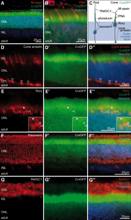
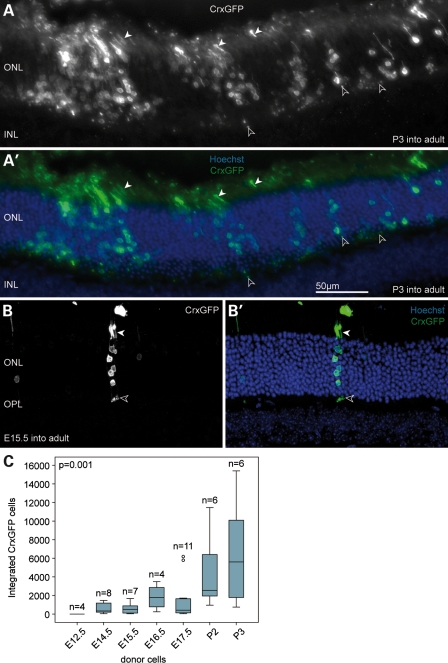
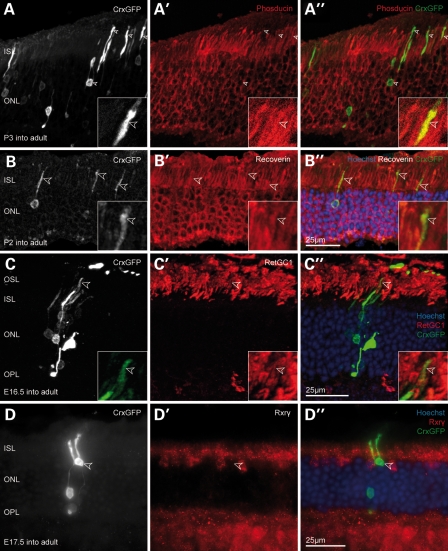
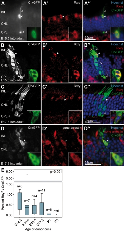
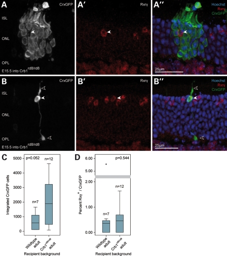
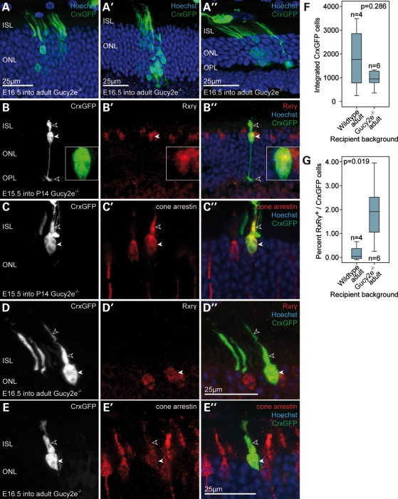
Retinal degenerative disease causing loss of photoreceptor cells is the leading cause of untreatable blindness in the developed world, with inherited degeneration affecting 1 in 3000 people. Visual acuity deteriorates rapidly once the cone photoreceptors die, as these cells provide daylight and colour vision. Here, in proof-of-principle experiments, we demonstrate the feasibility of cone photoreceptor transplantation into the wild-type and degenerating retina of two genetic models of Leber congenital amaurosis, the Crb1(rd8/rd8) and Gucy2e(-/-) mouse. Crx-expressing cells were flow-sorted from the developing retina of CrxGFP transgenic mice and transplanted into adult recipient retinae; CrxGFP is a marker of cone and rod photoreceptor commitment. Only the embryonic-stage Crx-positive donor cells integrated within the outer nuclear layer of the recipient and differentiated into new cones, whereas postnatal cells generated a 10-fold higher number of rods compared with embryonic-stage donors. New cone photoreceptors displayed unambiguous morphological cone features and expressed mature cone markers. Importantly, we found that the adult environment influences the number of integrating cones and favours rod integration. New cones and rods were observed in ratios similar to that of the host retina (1:35) even when the transplanted population consisted primarily of cone precursors. Cone integration efficiency was highest in the cone-deficient Gucy2e(-/-) retina suggesting that cone depletion creates a more optimal environment for cone transplantation. This is the first comprehensive study demonstrating the feasibility of cone transplantation into the adult retina. We conclude that flow-sorted embryonic-stage Crx-positive donor cells have the potential to replace lost cones, as well as rods, an important requirement for retinal disease therapy.


Figures

Figure 1.
Figure 1.
The CrxGFP transgene is expressed in both rod and cone photoreceptors. (A, B, DG) Immunostaining of CrxGFP adult retina. (D–G) Target protein immunostaining (red); (D′–G′) CrxGFP labelling (green); (D″–G″) three channels merge with Hoechst nuclear stain (blue). CrxGFP co-localizes with the cone photoreceptor markers, medium wavelength (M)-opsin (A), the lectin peanut agglutinin (PNA) (B), cone arrestin (D) and retinoid X receptor gamma (Rxrγ) (E). CrxGFP cells also co-label with the general mature photoreceptor markers recoverin (F) as well as retinal guanylate cyclase 1 (RetGC1/Gucy2e) (G). Double-labelled photoreceptors (D″, E″) exhibit characteristic cone morphologies with polymorphic nuclei near the outermost edge of the ONL (E, insets, open arrowheads) and large cone pedicles in the OPL (D, open arrowheads). (C) A cartoon representation of rod and cone photoreceptor morphology and marker expression. (A) is a confocal z-projection. ONL, outer nuclear layer; INL, inner nuclear layer; ISL, inner segment layer; OPL, outer plexiform layer.
Figure 2.
Figure 2.
The majority of transplanted CrxGFP cells develop into rod photoreceptors in the adult wild-type retina. (A) Clusters of transplanted P3 CrxGFP donor cells (green) migrated into the host ONL near the injection site. Cells exhibit typical rod photoreceptor morphology with highly condensed heterochromatin pattern, slender inner and outer segments (closed arrowheads) and spherical synapses (open arrowheads) in the OPL. (B) Transplanted E15.5 CrxGFP cells integrated into ONL and were usually observed as single- or small groups of photoreceptors. (C) Number of integrated CrxGFP cells per retina displayed in relationship to donor age. A trend was observed with significantly more cells integrating into the ONL with increased donor cell age (Spearman correlation, P < 0.01). Horizontal bars indicate the median value. (B) is a confocal z-projection. All retinae were examined 3 weeks post-transplantation. ONL, outer nuclear layer; INL, inner nuclear layer; OPL, outer plexiform layer. Hoechst nuclear stain (blue).
Figure 3.
Figure 3.
Transplanted CrxGFP cells with rod morphology co-label for functional photoreceptor markers. (A–D) CrxGFP labelling (grey scale), A′–D′ target protein immunostaining (red), A″–D″ three channels merge with CrxGFP (green) and Hoechst nuclear stain (blue). (A) Transplanted CrxGFP cells co-express the rod marker phosducin in inner segments and the cell body (arrowheads). Insets show a higher magnification and single optical section showing co-localization in the inner segment. (B) The general photoreceptor marker recoverin is expressed in transplanted CrxGFP cells. Arrowheads highlight inner segments. Insets show a higher magnification and single optical section showing co-localization in the inner segment. (C) Retinal guanylate cyclase 1 (RetGC1/Gucy2e) is expressed in the outer segments of transplanted rod photoreceptors (arrowheads). Insets display a higher magnification of the outer segment. (D) Transplanted cells with rod morphology do not co-label for the cone marker Rxrγ. All retinae were examined 3 weeks post-transplantation. (A–C) are confocal z-projections. ISL, inner segment layer; ONL, outer nuclear layer; OSL, outer segment layer; OPL, outer plexiform layer.
Figure 4.
Figure 4.
A small number of transplanted embryonic CrxGFP donor cells develop as cone photoreceptors in the wild-type retina. (A–D) A small number of transplanted embryonic CrxGFP precursors migrated into the host ONL from the sub-retinal space and differentiated into cone photoreceptors expressing the cone marker Rxrγ. Hoechst nuclear stain demarcates ONL in (A″–D″). Presumed cone cell bodies were without exception located at the outer aspect of the host ONL (solid arrowheads and insets), had large cone ribbon synapses (pedicles) in the OPL (open arrowheads) and exhibited a polymorphic heterochromatin structure typical for mature cone photoreceptors (Hoechst nuclear stain in insets). (A′–C′) Three examples of cone photoreceptors displaying nuclear Rxrγ immunostaining (solid arrowheads). (D) Example of a cone photoreceptor co-labelling with cone arrestin. (B″ and C″) Integrated cone cells (green; arrowheads) are located close to several transplanted rod cells (green, without arrowheads), which do not label with Rxrγ and have typical rod morphologies. (A–D) CrxGFP labelling (greyscale and inset green); (A′-D′) Rxrγ and cone arrestin immunostaining (red); (A″–D″) three channels merge with Hoechst nuclear stain (blue). All retinae were examined 3 weeks post-transplantation. (B–D) are confocal z-projections. (E) Graph of the efficiency of cone cell integration observed after transplants using CrxGFP donor cells at each donor age. Cone cell efficiency calculated as the proportion of newly integrated photoreceptors that are cones; Rxrγ CrxGFP double-positive cells/total CrxGFP. A trend was observed with significantly higher cone integration efficiencies observed using embryonic CrxGFP donor cells when compared with postnatal CrxGFP donor cells (Spearman correlation, P < 0.01). Horizontal bars indicate the median value. ISL, inner segment layer; ONL, outer nuclear layer; OPL, outer plexiform layer.
Figure 5.
Figure 5.
Transplantation of embryonic CrxGFP donor cells into the degenerating Crb1rd8/rd8 retina. (A) A cluster of transplanted CrxGFP rod photoreceptors (green) surrounding an Rxrγ-positive cone photoreceptor of the recipient retina (arrowhead). (B) Example of a transplanted CrxGFP cell exhibiting cone morphology (stubby inner segment, cone pedicle in the OPL, open arrowheads) and expressing the nuclear cone marker Rxrγ (solid arrowhead). (C) Comparison of the total number of integrated photoreceptors (rods and cones) after transplantation of E15.5 donor CrxGFP cells in the mutant and wild-type retina. Overall, photoreceptor integration was not significantly affected by the recipient environment (Mann–Whitney, P = 0.052). (D) Comparison of the cone integration efficiency (% Rxrγ/CrxGFP) of E15.5 CrxGFP cells in the mutant and wild-type retina. No statistically significant difference was observed (Mann–Whitney, P = 0.544). Horizontal bars indicate the median value. All retinae were examined 3 weeks post-transplantation. (A, B) are confocal z-projections. ISL, inner segment layer; ONL, outer nuclear layer; OPL, outer plexiform layer.
Figure 6.
Figure 6.
Transplantation of embryonic CrxGFP donor cells into the degenerating Gucy2e−/− retina. (A–E) Embryonic CrxGFP donor cells transplanted into the Gucy2e−/− degenerate retina at 2–3 months. (A) Transplanted CrxGFP donor cells exhibiting rod photoreceptor morphology after integrating into the ONL. (B–E) Examples of transplanted CrxGFP cells displaying cone morphology. (B,D) Cells express the nuclear cone marker Rxrγ (solid arrowheads and high magnification inset in B′). (C,E) Cone arrestin signal was observed in the inner segments and somata (open and solid arrowheads, respectively). (F) Comparison of the total number of integrated E16.5 CrxGFP photoreceptors (rods and cones) in the mutant and wild-type retina. Overall, photoreceptor integration was not significantly affected by the degenerative environment (Mann–Whitney, P = 0.286). Horizontal bars indicate the median value. All retinae were examined 3 weeks post-transplantation. (A–E) are confocal z-projections. (G) Comparison of the cone integration efficiency (% Rxrγ/CrxGFP) of E16.5 CrxGFP cells in the mutant and wild-type retina. Cone integration was significantly more efficient after CrxGFP donor cell transplantation into the degenerating Gucy2e−/− retina when compared with wild-type (Mann–Whitney, P = 0.019). ISL, inner segment layer; ONL, outer nuclear layer; OPL, outer plexiform layer.

Similar articles

Cited by

References

    1. Morrow E.M., Furukawa T., Cepko C.L. Vertebrate photoreceptor cell development and disease. Trends Cell Biol. 1998;8:353–358. doi:10.1016/S0962-8924(98)01341-5. - DOI - PubMed
    1. Hartong D.T., Berson E.L., Dryja T.P. Retinitis pigmentosa. Lancet. 2006;368:1795–1809. doi:10.1016/S0140-6736(06)69740-7. - DOI - PubMed
    1. Lamba D.A., Karl M.O., Reh T.A. Strategies for retinal repair: cell replacement and regeneration. Prog. Brain Res. 2009;175:23–31. doi:10.1016/S0079-6123(09)17502-7. - DOI - PubMed
    1. West E.L., Pearson R.A., MacLaren R.E., Sowden J.C., Ali R.R. Cell transplantation strategies for retinal repair. Prog. Brain Res. 2009;175:3–21. doi:10.1016/S0079-6123(09)17501-5. - DOI - PMC - PubMed
    1. MacLaren R.E., Pearson R.A., MacNeil A., Douglas R.H., Salt T.E., Akimoto M., Swaroop A., Sowden J.C., Ali R.R. Retinal repair by transplantation of photoreceptor precursors. Nature. 2006;444:203–207. doi:10.1038/nature05161. - DOI - PubMed

Publication types