Review
. 2010 Dec;22(6):687-95.
doi: 10.1097/MOP.0b013e32833f56d5.

Holoprosencephaly: recommendations for diagnosis and management

Affiliations
Review

Holoprosencephaly: recommendations for diagnosis and management

Emily F Kauvar et al. Curr Opin Pediatr. 2010 Dec.

Abstract

Purpose of review: This review presents recent advances in our understanding and clinical management of holoprosencephaly (HPE). HPE is the most common developmental disorder of the human forebrain and involves incomplete or failed separation of the cerebral hemispheres. The epidemiology, clinical features, causes, diagnostic approach, management, and outcomes of HPE are discussed.

Recent findings: Chromosomal abnormalities account for the most commonly identified cause of HPE. However, there are often unidentifiable causes in patients with nonsyndromic, nonchromosomal forms of HPE. The prevalence of HPE may be underestimated given that patients with mild forms often are not diagnosed until they present with severely affected children. Pregestational maternal diabetes mellitus is the most recognized risk factor for HPE, as supported by recent large-scale epidemiological studies. Genetic studies using microarray-based comparative genomic hybridization technology have resulted in better characterization of important HPE loci.

Summary: HPE encompasses a wide spectrum of forebrain and midline defects, with an accompanying wide spectrum of clinical manifestations. A coordinated, multidisciplinary care team is required for clinical management of this complex disorder. Further research will enable us to better understand the pathogenesis and causes of HPE, and thus to improve the genetic counseling of patients and their families.


Figures
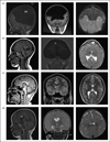
Figure 1
Figure 1. The neurological spectrum of holoprosencephaly
Sagittal, coronal, and axial MRI images (left to right, respectively) depict the spectrum of HPE, from the most severe form, alobar HPE (a), to the mildest form middle interhemispheric variant (MIHV) HPE (d). (a) MRI of a 10-month-old infant with alobar HPE that shows entire hemispheric fusion, an absent falx cerebri, an absent corpus callosum, a monoventricle, a large dorsal cyst (DC) with massive hydrocephaly, and a central gray mass that consists of poorly differentiated caudate and lentiform nuclei fused in the midline (white arrows). (b) MRI of an 8-month-old infant with semilobar HPE that shows anterior hemispheric fusion (curved white arrow), a present posterior falx cerebri, absent frontal horns (curved black arrow), and absence of the genu of the corpus callosum (black arrow), but presence of the splenium and some of the posterior body (white arrow). (c) MRI of an 18-year-old individual with lobar HPE that shows fusion of the deep mesial and basal frontal lobes (superior curved white arrow) and continuous gray matter in the basal frontal region (inferior curved white arrow), a full falx cerebri, a small frontal horn (black arrow), an absent head of the corpus callosum with the body and splenium present (white arrow), and separation of the caudate and lentiform nuclei (curved black arrow). (d) MRI of a 10-month-old infant with MIHV HPE that shows midfrontal hemispheric fusion (curved black arrow) and an absent body of the corpus callosum that puckers superiorly due to cortical fusion (white arrow). Images courtesy of Jin S. Hahn, MD of Stanford University School of Medicine, Lucile Packard Children’s Hospital, and Stanford Carter Center for Brain Research in Holoprosencephaly and Related Malformations.
Figure 2
Figure 2. The craniofacial spectrum of holoprosencephaly
The spectrum of HPE ranges from the most severe form, alobar HPE (a), to the mildest form, microform HPE (d) (left to right, respectively). (a, b) Two patients with alobar HPE who have different degrees of craniofacial involvement: (a) a patient with synophthalmia (two eyes fused) and a proboscis (nose-like structure); (b) a patient with hypotelorism, a flat nasal bridge, colobomata, and a facial cleft; (c) a patient with lobar HPE, who has hypotelorism and a flat nasal bridge; (d) a patient with microform HPE, who has hypotelorism and a single maxillary central incisor. Images modified and reproduced with permission from the following: (a,d) from [9], Nature Publishing Group; (b) from [10], Springer Science+Business Media, Fig. 1c; and (c) from [11••], BMJ Publishing Group, Ltd.
Figure 3
Figure 3. The diagnostic evaluation and clinical management of children with holoprosencephaly
The illustrated flowchart summarizes the recommended approach for the diagnostic evaluation and clinical management of children with HPE. *See Pineda-Alvarez et al. [36•] for further details on the molecular evaluation. **See Levey et al. [3••] for further details on clinical problems and management. Clinical work-up and management should be guided by the primary care physician and appropriate consultative specialties. The above list is not intended to be exhaustive or comprehensive, and it may not be applicable to all patients. ACTH, adrenocorticotropic hormone; CSF, cerebrospinal fluid; DDAVP, desmopressin acetate; DI, diabetes insipidus; GH, growth hormone; ITB, intrathecal baclofen; PT, physical therapy; OT, occupational therapy; SLP, speech/language pathology; TH, thyroid hormone.

References

    1. Shiota K, Yamada S. Early pathogenesis of holoprosencephaly. Am J Med Genet C Semin Med Genet. 2010;154C:22–28. - PubMed
    1. Orioli IM, Castilla EE. Epidemiology of holoprosencephaly: prevalence and risk factors. Am J Med Genet C Semin Med Genet. 2010;154C:13–21. - PubMed
    1. Levey EB, Stashinko E, Clegg NJ, Delgado MR. Management of children with holoprosencephaly. Am J Med Genet C Semin Med Genet. 2010;154C:183–190. The authors of this article are clinicians and researchers in the Carter Centers for Brain Research in Holoprosencephaly and Related Malformations. This article provides a very thorough and valuable resource for clinicians who care for children with HPE as it reviews the clinical management and outcomes of children with HPE.

    1. Solomon BD, Pineda-Alvarez DE, Mercier S, et al. Holoprosencephaly flashcards: a summary for the clinician. Am J Med Genet C Semin Med Genet. 2010;154C:3–7. This article provides a summary for clinicians of key information about HPE, in the format of ‘flashcards’. The flashcards illustrate the prevalence, causes, and types of HPE, as well as the craniofacial and neurological findings, physical examination features, the clinical approach, and resources.

    1. Hahn JS, Barnes PD. Neuroimaging advances in holoprosencephaly: refining the spectrum of the midline malformation. Am J Med Genet C Semin Med Genet. 2010;154C:120–132. This article is a valuable resource for understanding the spectrum of HPE specifically as it relates to neuroimaging. The article thoroughly describes the cerebral structure of each type of HPE, with accompanying neuroimages and labels to highlight key features.

Publication types