. 2011 Jan;19(1):36-45.
doi: 10.1038/mt.2010.205. Epub 2010 Sep 21.

Gene therapy of mdx mice with large truncated dystrophins generated by recombination using rAAV6

Affiliations

Gene therapy of mdx mice with large truncated dystrophins generated by recombination using rAAV6

Guy L Odom et al. Mol Ther. 2011 Jan.

Abstract

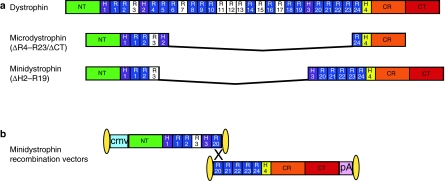
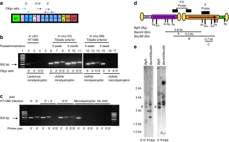
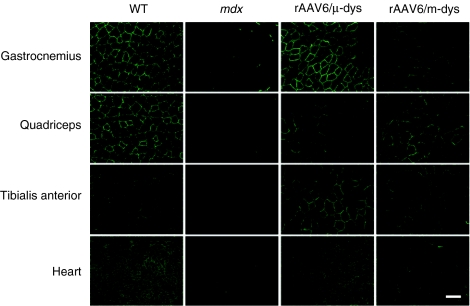
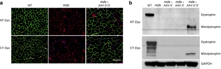
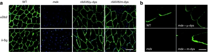
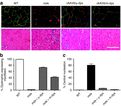
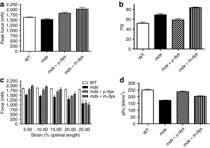
Recombinant adeno-associated viral (rAAV) vector-mediated gene transfer represents a promising approach for many diseases. However, the applicability of rAAV vectors has long been hindered by the small (~4.8 kb) DNA packaging capacity. This limitation can hamper the packaging and delivery of critical regulatory elements and/or larger coding sequences, such as the ~14-kb dystrophin complementary DNA (cDNA) that is of interest for gene therapy of Duchenne muscular dystrophy (DMD). Here, we have demonstrated reconstitution of an expression cassette (7.3 kb) encoding a highly functional "minidystrophin" protein (ΔH2-R19, 222 kd) in vivo following intravascular co-delivery of two independent rAAV6 vectors sharing a central homologous recombinogenic region of 372 nucleotides. Similar to previously reported trans-splicing approaches, one rAAV vector provides the promoter with the ~1/2 initial portion of minidystrophin, while the second vector provides the remaining minidystrophin cDNA followed by the polyadenylation signal. Significantly, administering a modest dose [2 × 10(12) vector genomes (vg)] of the two minidystrophin-encoding rAAV vectors to dystrophic mice elicited an improvement of physiological performance indicative of prevention or amelioration of the disease state. These studies provide evidence that functional dystrophin transgenes larger than that typically carried by a single rAAV genome can be reconstituted in vivo by homologous recombination (HR) following intravascular co-delivery with rAAV6.


Figures

Figure 1
Figure 1
Schematic representation of rAAV6 recombination vectors for generation of the 222 kd minidystrophin protein. (a) Structural domains of the full-length dystrophin, microdystrophin (ΔR4–R23/ΔCT), and minidystrophin (ΔH2–R19) proteins. (b) The rAAV6 recombination vectors are flanked by the inverted terminal repeats (ITRs) of serotype 2 at both ends (oblong circles). The locations of the CMV promoter (blue) and SV40 polyadenylation (pA) signal (pink) sequences are shown. The two vectors encode either the 5′ or the 3′ approximate half portions of the minidystrophin transgene. The 5′ cassette ends within exon 53 (approximate middle of R21) at nucleotide 7,980 of the human dystrophin cDNA sequence. The 3′ cassette begins within exon 51 (approximate middle of H3) at nucleotide 7,608 and terminates within exon 79 at nucleotide 11,564 of the human dystrophin complementary DNA sequence. Following coadministration in vivo, the two vectors assemble through a homologous recombination mechanism (×), allowing formation of an mRNA that encodes a larger and nearly fully functional minidystrophin where the correctly recombined genome is ~7.3 kb. Note that basic repeats are shaded white. CMV, cytomegalovirus; CR, cysteine-rich domain; CT, carboxy-terminal domain; H1–4, hinges 1–4; NT, amino-terminus; R1–24; spectrin-like repeats 1–24.
Figure 2
Figure 2
Molecular analysis of rAAV6/minidystrophin genomes. (a) Oligonucleotide primer pairs used for the detection of individual rAAV6/minidystrophin vector genomes (vg). The 5′oligo pair (green arrows) was designed to amplify the 5′ rAAV genome in the region encoding spectrin-like repeats 1–3 (R1–R3; see below); the 3′ oligo pair (blue arrows) amplifies the 3′ rAAV genome in the region encoding R21–R22; the 5′/3′oligo pair (red arrows) yields a product representative of the recombination junction, amplifying DNA encoding the region from H3–R21. (b) PCR analysis of vg using DNA extracted from the tibialis anterior (TA) muscles of mice administered the dual recombination vectors intravenously (i.v.; 2 × 10¹² vg, lanes 6–11) or intramuscularly (i.m.; 1 × 10¹⁰ vg, lanes 12–14). As a negative control, some mice were also injected with the single rAAV6/microdystrophin vector (i.m.; 1 × 10¹⁰ vg, lanes 15–17). As a positive control, HT1080 cells were infected at a multiplicity of infection of 10 with a lentiviral vector expressing minidystrophin (lanes 2–4). (c) PCR analysis of vg obtained via DNA extracted from HT1080 cells infected with individual or dual rAAV-minidystrophin overlapping vectors. When mixing DNA obtained from cells infected independently with either AAV-5′ or AAV-3′ vectors (lanes marked 5′ + 3′) the 5′/3′ primer pair (lane 6) resulted in no amplification (i.e. no bridged, or recombinant PCR products were detected). Amplification of the 5′/3′ region was only detected when the two vectors were coinfected into the cells (lanes marked 5′/3′). (d) Diagram of rAAV6/minidystrophin recombination vg with restriction enzyme cut sites and representative probes used for Southern blot analysis as described for e. Dual-vector head-to-tail orientations are represented by arrows pointing in the same direction. Inverted terminal repeats are shown as oblong circles. The vector–vector junction may contain 0, 1, or 2 ITRs, and as a result the predicted sizes of hybridized genomes (Supplementary Table S1) are accurate to within ~160 base pairs. The probes utilized hybridize to sites indicated by the black bars shown above the vector genome diagram. (e) Southern blot analysis of total DNA from mdx4cv TA muscles harvested 5 months after i.v. injection of the dual rAAV6/minidystrophin recombination vectors. DNA was digested with BglII or in a separate reaction with BamHI plus Bsu36I followed by hybridization with each of the two probes as indicated. The positions of size standards (in kilobases) are shown on the left. In myofibers transduced with both of the minidystrophin vectors, the BamHI/Bsu36I-digested DNA, when hybridized with the 5′/3′ probe, displayed a band of 2.3 kb in size representing reconstituted minidystrophinΔR4–R23/ΔCT genomes (a). Similarly the BglII-digested DNA when hybridized with either the 5′/3′ or the 3′ probe revealed a 3.3-kb band, diagnostic for the 5′/3′—minidystrophin genomes arranged head–tail (b). The 3′ probe also revealed a 0.7-kb band representing total 3′-minidystrophin monomeric genomes as a result of detecting the BamHI-digested DNA (c). CMV, cytomegalovirus; rAAV, recombinant adeno-associated virus.
Figure 3
Figure 3
Mini- and microdystrophin generated in vivo following delivery of rAAV6 vectors both properly localized to the sarcolemma of striated muscles in treated mdx mice. Administration consisted of injecting 2 × 10¹² vector genomes i.v. in a 0.3 ml total volume. Analysis was at 4 months postinjection. Green fluorescence represents dystrophin staining as detected by a polyclonal antibody to the amino-terminal domain. Bar = 100 µm. rAAV, recombinant adeno-associated virus; WT, wild type.
Figure 4
Figure 4
Minidystrophin protein expression detection as revealed by immunofluorescence and immunoblotting using either an N- (NT) or C-terminal (CT) dystrophin antibody. (a) Cryosections of tibialis anterior muscle treated with rAAV-minidystrophin overlapping vectors demonstrate proper localization at the sarcolemma when detected by NT or CT antidystrophin antibody. (b) Similarly immunoblotting of whole muscle lysates from tibialis anterior muscle demonstrate detection of the 222 kd protein with either the NT or CT antidystrophin antibody. rAAV, recombinant adeno-associated virus; WT, wild type.
Figure 5
Figure 5
Intravenous delivery of rAAV6/ microdystrophin or dual rAAV6/minidystrophin recombination vectors results in the proper localization of cytoplasmic peripheral and integral membrane protein components of the dystrophin–glycoprotein complex and concentration at the myotendinous junctions of treated mdx mice. (a) TA muscles from treated and untreated mice were cryosectioned followed by immunofluorescence staining for α-dystrobrevin-2 (αDb2) and δ-sarcoglycan (δSg), shown as green labeling, with nuclei seen as blue. Bar = 100 µm. (b) An amino-terminal dystrophin antibody was used for immunofluorescent staining of longitudinally cryosectioned gastrocnemius muscles from wild-type, mdx4cv, and mdx4cv treated with either rAAV6/ microdystrophin or rAAV6/ minidystrophin recombination vectors. Bar = 40 µm. rAAV, recombinant adeno-associated virus; WT, wild type.
Figure 6
Figure 6
rAAV6-mediated transduction with minidystrophin improves muscle histopathology in mdx mice. (a) Upper panel shows immunofluorescent staining of tibialis anterior muscle for B2 laminin (red), dystrophin (green), and nuclei (blue). The lower panel shows adjacent sections stained with hematoxylin and eosin. (b) Shows the percentage of dystrophin-positive fibers in the different experimental groups while c shows the percentage of myofibers with centrally located nuclei in the TA muscles. Bar = 100 µm. Bars represent SEM. rAAV, recombinant adeno-associated virus; WT, wild type.
Figure 7
Figure 7
Intravenous administration of rAAV6/minidystrophin recombination vectors to mdx mice results in increased muscle function. In comparison to mdx4cv tibialis anterior (TA) muscles expressing minidystrophin, the TA muscles of untreated mdx4cv mice exhibited (a) reduced force generating capacity; (b) decreased mass; (c) an increased susceptibility to eccentric contraction-induced injury, and (d) decreased force generation per cross-sectional area (specific force). Bars represent the SEM. sPo, specific force; WT, wild type.

Similar articles

Cited by

References

    1. Berns KI. Parvovirus replication. Microbiol Rev. 1990;54:316–329. - PMC - PubMed
    1. Chiorini JA, Wiener SM, Owens RA, Kyöstió SR, Kotin RM., and, Safer B. Sequence requirements for stable binding and function of Rep68 on the adeno-associated virus type 2 inverted terminal repeats. J Virol. 1994;68:7448–7457. - PMC - PubMed
    1. Ryan JH, Zolotukhin S., and, Muzyczka N. Sequence requirements for binding of Rep68 to the adeno-associated virus terminal repeats. J Virol. 1996;70:1542–1553. - PMC - PubMed
    1. Snyder RO, Im DS, Ni T, Xiao X, Samulski RJ., and, Muzyczka N. Features of the adeno-associated virus origin involved in substrate recognition by the viral Rep protein. J Virol. 1993;67:6096–6104. - PMC - PubMed
    1. Muzyczka N. Use of adeno-associated virus as a general transduction vector for mammalian cells. Curr Top Microbiol Immunol. 1992;158:97–129. - PubMed

Publication types

MeSH terms