

doi: 10.1002/adma.201001525.
Nano-layered microneedles for transcutaneous delivery of polymer nanoparticles and plasmid DNA
Affiliations
- PMID: 20859938
- PMCID: PMC3030257
- DOI: 10.1002/adma.201001525
Item in Clipboard
Nano-layered microneedles for transcutaneous delivery of polymer nanoparticles and plasmid DNA
Adv Mater.
.
No abstract available
Figures
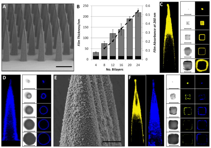
(A) SEM micrograph of uncoated PLGA microneedle arrays of pyramidal geometry (scale - 500μm). (B) Film growth (left axis) and absorbance (right axis) for (Poly-1/pLUC)n multilayers assembled on silicon/quartz substrates bearing a (PS/SPS)20 initiating layer (black bar - (PS/SPS)20, grey bar - (Poly-1/pLUC)n, dashed line – Ab-260 nm). (C, D) Representative confocal micrographs showing a (C) (PS/SPS)20-(Poly-1/Cy3-pLUC)24 coated microneedle and a (D) (PS/SPS)20-(Poly-1/DiI-PLGA NP)4 coated microneedle (left – transverse section, right – lateral sections, 200μm intervals, scale - 200 μm). (E) SEM micrograph showing a (PS/SPS)20-(Poly-1/PLGA NP)4 coated microneedle array (scale - 50μm). (F) Representative confocal micrographs showing a (PS/SPS)20-(Poly-1/Cy3-pLUC)24-(Poly-1/DiD-PLGA NP)4 co-coated microneedle (transverse and lateral sections, left – Cy3-pLUC, right – DiD-PLGA NP, 200μm intervals, scale - 200 μm).
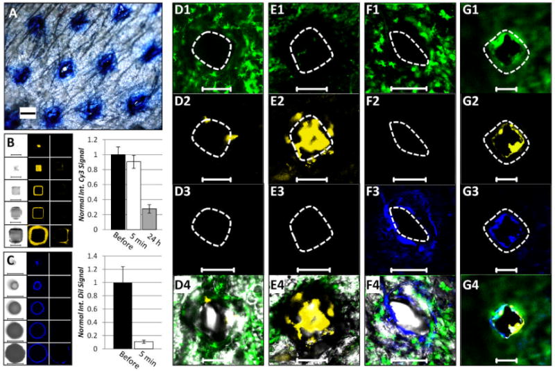
(A) Optical micrograph of ear skin showing microneedle penetration pattern stained using trypan blue (scale – 100μm). (B) Representative confocal z-stacks and quantification (n = 6) of (PS/SPS)20-(Poly-1/Cy3-pLUC)24-coated microneedle arrays (left – brightfield, middle – before application, right – after 24 hour application, 200μm interval, scale - 200μm). (C) Representative confocal z-stacks and quantification (n = 6) of (PS/SPS)20-(Poly-1/DiI-PLGA-NP)4 coated microneedle arrays (left – brightfield, middle – before application, right – after 5 minute application, 200μm interval, scale – 200μm). Representative confocal micrographs (1 – MHC-GFP II, 2 – Cy3-pLUC, 3 – DiI/D-PLGA NP, 4 – overlay, scale – 200μm) showing dorsal ear skin following (D) 5 minute and (E) 24 hour application of a (PS/SPS)20-(Poly-1/Cy3-pLUC)24 coated microneedle array, (F) 5 minute (PS/SPS)20-(Poly-1/DiI-PLGA-NP)4 coated microneedle application, and (G) 24 hour (PS/SPS)20-(Poly-1/Cy3-pLUC)24-(Poly-1/DiD-PLGA NP)4 coated microneedle application.
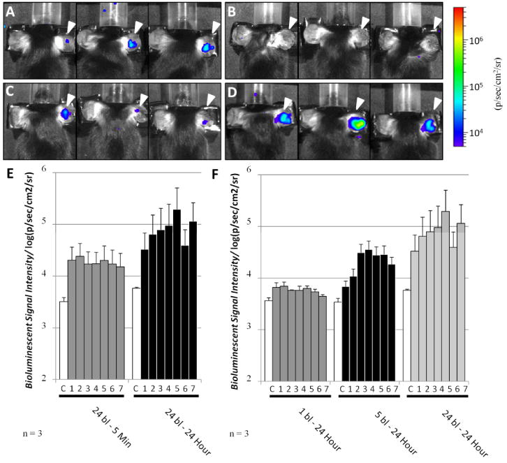
In vivo bioluminescent signal observed in C57BL/6 mice (n = 3) following treatment with a (PS/SPS)20-(Poly-1/pLUC)n – coated microneedle array to the right ear (denoted by arrow): (A) 24 bilayers for 5 minutes, (B) 1 bilayer for 24 hours, (C) 5 bilayers for 24 hours, and (D) 24 bilayers for 24 hours. The bioluminescent results following treatment are summarize in (E, F) for 7 days together with the negative control signal (denoted C) collected from the untreated ear, with (E) demonstrating the effect of application time and (F) showing the result of increasing pLUC dosage.
Similar articles
-
Immunostimulatory effect of tetanus toxoid loaded chitosan nanoparticles following microneedles assisted immunization.Nanomedicine. 2016 Jan;12(1):213-22. doi: 10.1016/j.nano.2015.10.009. Epub 2015 Nov 10. Nanomedicine. 2016. PMID: 26554391
-
In vitro and in vivo characterization of MEMS microneedles.Biomed Microdevices. 2005 Mar;7(1):47-52. doi: 10.1007/s10544-005-6171-y. Biomed Microdevices. 2005. PMID: 15834520
-
Multifunctional nanorods for gene delivery.Nat Mater. 2003 Oct;2(10):668-71. doi: 10.1038/nmat974. Epub 2003 Sep 14. Nat Mater. 2003. PMID: 12970757
-
Single-cell manipulation and DNA delivery technology using atomic force microscopy and nanoneedle.J Nanosci Nanotechnol. 2014 Jan;14(1):57-70. doi: 10.1166/jnn.2014.9115. J Nanosci Nanotechnol. 2014. PMID: 24730251 Review.
-
[Application of MEMS microneedles array in biomedicine].Sheng Wu Yi Xue Gong Cheng Xue Za Zhi. 2004 Jun;21(3):482-5. Sheng Wu Yi Xue Gong Cheng Xue Za Zhi. 2004. PMID: 15250162 Review. Chinese.
Cited by
-
Oxidation-Responsive, Tunable Growth Factor Delivery from Polyelectrolyte-Coated Implants.Adv Healthc Mater. 2021 May;10(9):e2001941. doi: 10.1002/adhm.202001941. Epub 2021 Mar 18. Adv Healthc Mater. 2021. PMID: 33738985 Free PMC article.
-
Electrospun nanofibers as versatile interfaces for efficient gene delivery.J Biol Eng. 2014 Dec 9;8:30. doi: 10.1186/1754-1611-8-30. eCollection 2014. J Biol Eng. 2014. PMID: 25926887 Free PMC article. Review.
-
Recent Advances in Hybrid Biomimetic Polymer-Based Films: from Assembly to Applications.Polymers (Basel). 2020 Apr 26;12(5):1003. doi: 10.3390/polym12051003. Polymers (Basel). 2020. PMID: 32357541 Free PMC article. Review.
-
Releasable layer-by-layer assembly of stabilized lipid nanocapsules on microneedles for enhanced transcutaneous vaccine delivery.ACS Nano. 2012 Sep 25;6(9):8041-51. doi: 10.1021/nn302639r. Epub 2012 Aug 30. ACS Nano. 2012. PMID: 22920601 Free PMC article.
-
Controlling the surface-mediated release of DNA using 'mixed multilayers'.Bioeng Transl Med. 2016 Jun;1(2):181-192. doi: 10.1002/btm2.10023. Epub 2016 Aug 26. Bioeng Transl Med. 2016. PMID: 27981243 Free PMC article.
References
Publication types
MeSH terms
Substances
Grants and funding
LinkOut - more resources
Full Text Sources
Other Literature Sources
