Review
. 2011 Mar-Apr;3(2):162-73.
doi: 10.1002/wnan.116. Epub 2010 Sep 21.

Revisiting an old friend: manganese-based MRI contrast agents

Affiliations
Review

Revisiting an old friend: manganese-based MRI contrast agents

Dipanjan Pan et al. Wiley Interdiscip Rev Nanomed Nanobiotechnol. 2011 Mar-Apr.

Abstract

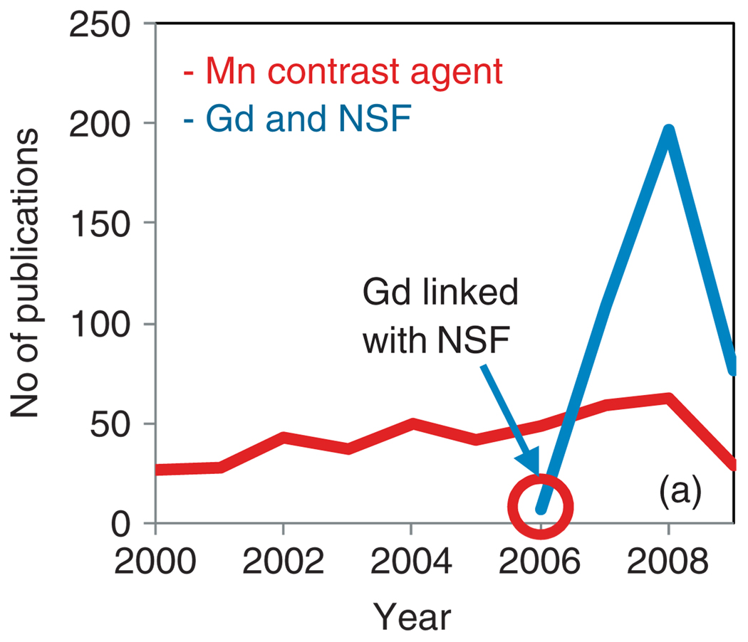
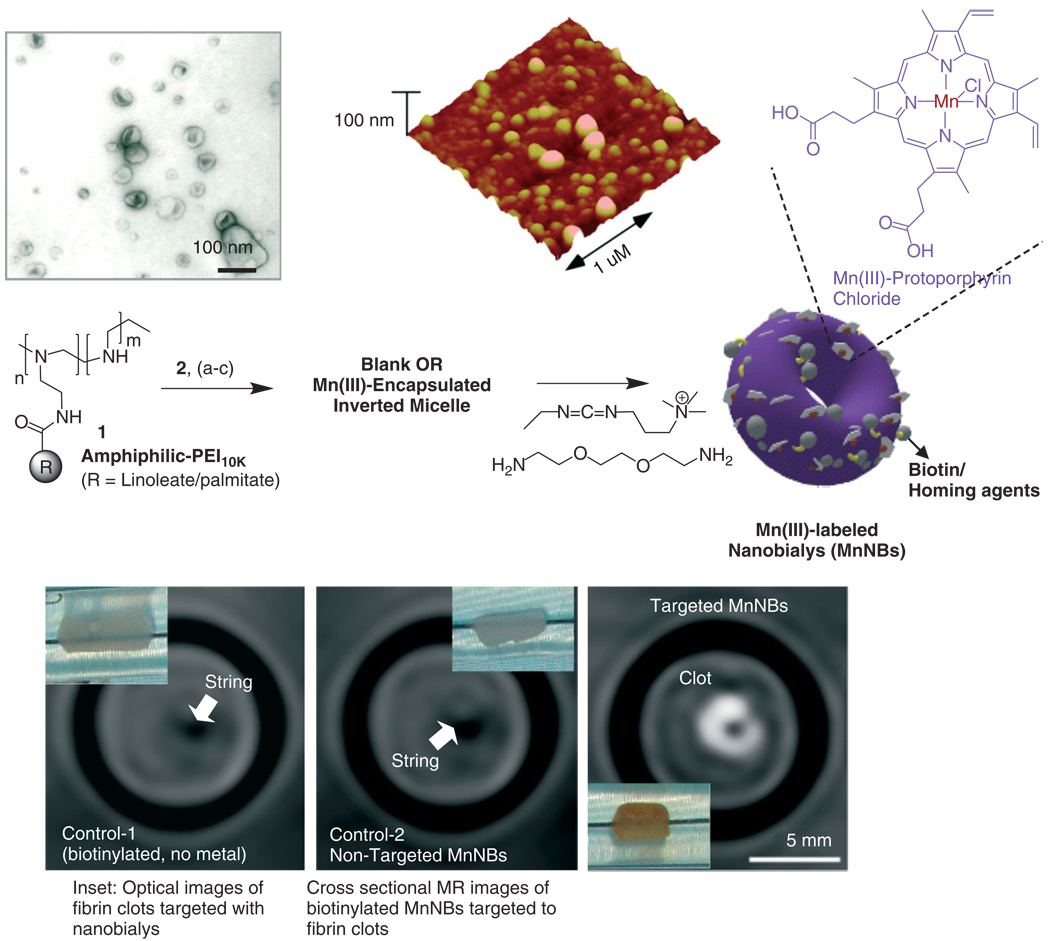
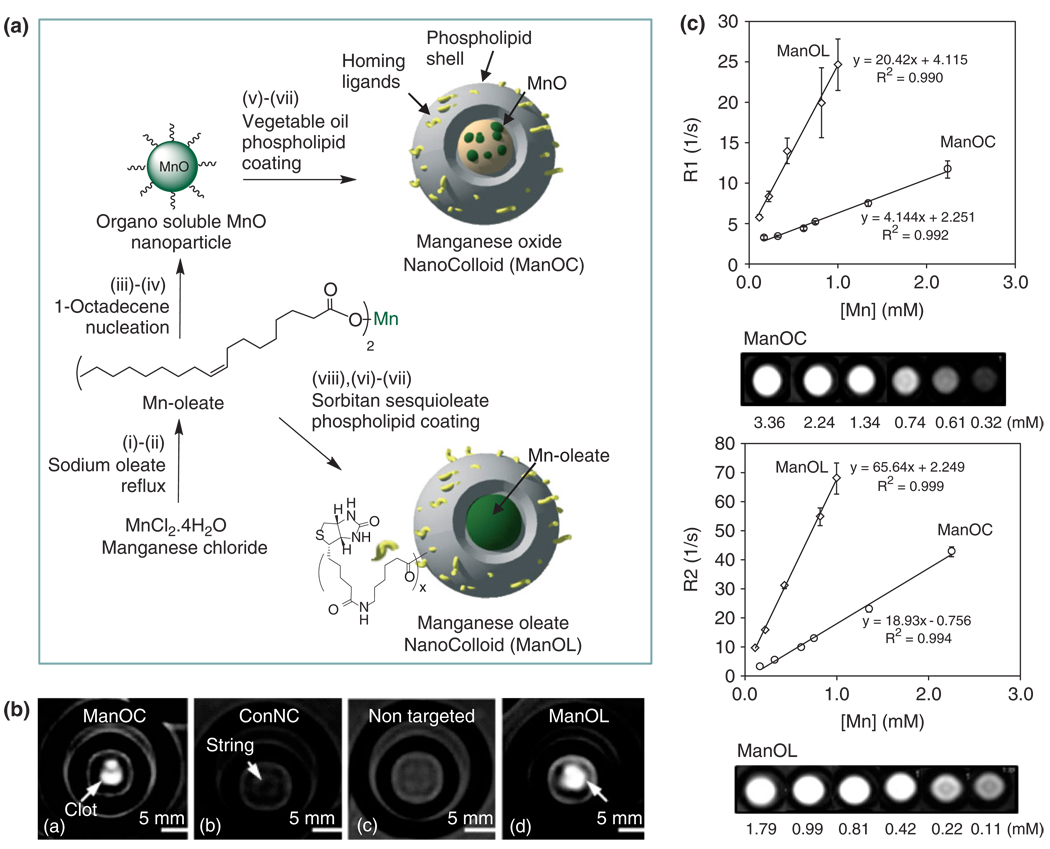
Non-invasive cellular and molecular imaging techniques are emerging as a multidisciplinary field that offers promise in understanding the components, processes, dynamics and therapies of disease at a molecular level. Magnetic resonance imaging (MRI) is an attractive technique due to the absence of radiation and high spatial resolution which makes it advantageous over techniques involving radioisotopes. Typically paramagnetic and superparamagnetic metals are used as contrast materials for MR based techniques. Gadolinium has been the predominant paramagnetic contrast metal until the discovery and association of the metal with nephrogenic systemic fibrosis (NSF) in some patients with severe renal or kidney disease. Manganese was one of the earliest reported examples of paramagnetic contrast material for MRI because of its efficient positive contrast enhancement. In this review manganese based contrast agent approaches will be presented with a particular emphasis on nanoparticulate agents. We have discussed both classically used small molecule based blood pool contrast agents and recently developed innovative nanoparticle-based strategies highlighting a number of successful molecular imaging examples.


Figures

FIGURE 1
FIGURE 1
Publication trend from Scopus: surge in publication related to Gd and NSF.
FIGURE 2
FIGURE 2
Manganese-based contrast agent families. Inset: Structure of dipyridoxal diphosphate (DPDP), the clinically approved manganese (II) agent for hepatobiliary imaging.
FIGURE 3
FIGURE 3
Structures of polycarboxylates and porphyrins used as ligands for manganese.
FIGURE 4
FIGURE 4
Example of convertible inorganic contrast agents.
FIGURE 5
FIGURE 5
Surface attached manganese-oxo clusters improve MR contrast (PS: polystyrene beads).
FIGURE 6
FIGURE 6
Preparation and characterization of manganese nanobialys: (Top left) TEM (drop deposited over nickel grid, 1% uranyl acetate) and (top right) atomic force microscope (AFM) image of nanobialys (drop deposited over glass) Reaction conditions: (1) anhydrous chloroform, gentle vortexing, room temperature; (2) aqueous solution of 2 [Mn(III)-protoporphyrin], inversion, room temperature, filter using short bed of sodium sulfate and cotton; (3) Biotin-Caproyl-PE, filter mixed organic solution using cotton bed, 0.2 µM water, vortex, gently evaporation of chloroform at 45°C, 420 mbar, 0.2 µM water, sonic bath, 50°C, 1/2 h, dialysis (2 kDa MWCO cellulose membrane) against water. (Bottom) MRI images of fibrin-targeted nanobialys (right) or control nanoparticles (bottom left) and (bottom middle) bound to cylindrical plasma clots measured at 3 T.
FIGURE 7
FIGURE 7
Preparation and characterization of manganese nanocolloids: Reaction conditions: (A) Preparation of ManOC and ManOL. (i)–(ii) sodium oleate, reflux, stirring; (iii)–(iv) 1-octadecene, 325°C/70 min, stirring; (v) suspended with vegetable oil (2 w/v%), vortex, mixing; evaporation of chloroform under reduced pressure, 45°C; (vi) thin film formation from phospholipids mixture; (vii) homogenization, 20,000 psi, 4 min, 0°C; (viii) Mn-oleate, suspended with sorbitan sesquioleate (>2 w/v%), vortex, mixing, evaporation of chloroform under reduced pressure, 45°C; then steps (vi), followed by (vii). (B) MRI images of fibrin-targeted nanocolloids: (a) ManOC; (b) ConNC; (c) nontargeted-ManOL and (d) ManOL, bound to cylindrical plasma clots measured at 3 T (pixel dimension: 0.73 mm × 0.73 mm × 5 mm slice thickness). (C) MR characterization of ManOC and ManOL in suspension: (top) ionic R1 and (bottom) R2 relaxivity. The measured R1 relaxation rate at 3 T for ManOC (circles) and ManOL (diamonds) as a function of manganese concentration. (Reproduced with permission from Ref . Copyright 2009 American Chemical Society).

References

    1. Pan D, Lanza GM, Wickline SA, Caruthers SD. Nanomedicine: perspective and promises with ligand-directed molecular imaging. Eur J Radiol. 2009;70:274–285. - PubMed
    1. Pan D, Caruthers SD, Chen J, Winter PM, Senpan A, Schmieder AH, Wickline SA, Lanza GM. Nanomedicine strategies for molecular targets with MRI and optical imaging. Future Med Chem. 2010;2:471–491. - PMC - PubMed
    1. Massoud TF, Gambhir SS. Molecular imaging in living subjects: seeing fundamental biological processes in a new light. Genes Dev. 2003;17:545–580. - PubMed
    1. Chan KW, Wong W-T. Smallmolecular gadolinium(III) complexes as MRI contrast agents for diagnostic imaging. Coord Chem Rev. 2007;17:2428–2451.
    1. Sieber MA, Steger-Hartmann T, Lengsfeld P, Pietsch H. Gadolinium-based contrast agents and NSF: evidence from animal experience. J Magn Reson Imaging. 2009;30:1268–1276. - PubMed