

An important Norwegian contribution to the study of the bursae of the upper and lower extremities
- PMID: 20860444
- PMCID: PMC3214749
- DOI: 10.3109/17453674.2010.506633
An important Norwegian contribution to the study of the bursae of the upper and lower extremities
Abstract
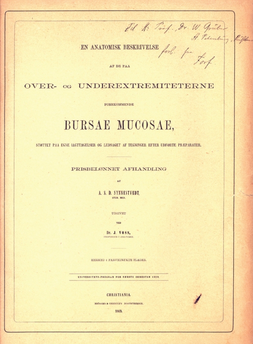
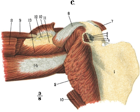
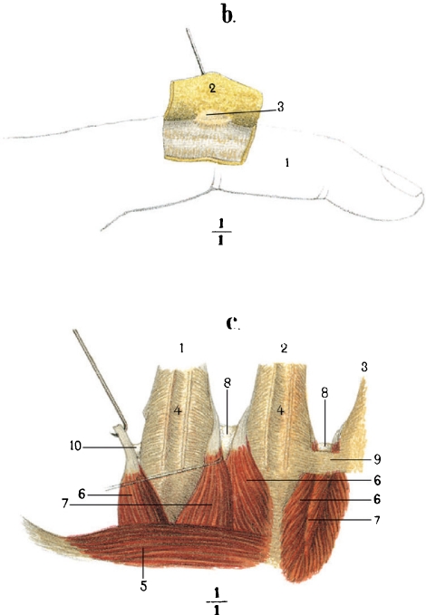
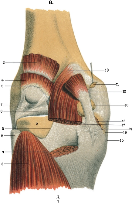
We present a critical analysis of the monograph of A.S.D. Synnestvedt (1869) “En anatomisk beskrivelse af de paa over- og underestremiteterne forekommende Bursae mucosae”. The analysis was completed using anatomical information from the historically oldest publications dealing with the bursae of the extremities: Albinus (1734) , Monro (1788) , Rosenmüller (1799) . We are of the opinion that Synnestvedt's publication is important, not only historically but also as a source of information for recent medical practitioners. Synnestvedt's monograph has a wealth of literary citations, unambiguous opinions of seasoned anatomists regarding the structure and function of the synovial membrane, and detailed descriptions of dissections he performed on fetal and adult cadavers. The information in this publication may enhance the diagnosis of bursopathies and enthesopathies of the extremities.
Figures

References
-
- Albert E. Achillodynie. Wiener Mediz Presse. 1893;34:43, 4.
-
- Albinus BS. Historia musculorum hominis. 1734. Th. Haak & H. Mulhovium Leidae Batavorum.
-
- Donath T. Pergamon Press; Oxford: 1969. Anatomical dictionary with nomenclatures and explanatory notes.
-
- FCAT . Thieme: Stuttgart; 1998. Terminologia Anatomica.
-
- University Press; Glasgow: 1933. Final Report of the Committee appointed by the Anatomical Society of Great Britain and Ireland on June 22nd, 1928.
Publication types
MeSH terms
Personal name as subject
- Actions
LinkOut - more resources
Full Text Sources
Medical
Research Materials
Miscellaneous