. 2010 Oct;101(10):2128-36.
doi: 10.1111/j.1349-7006.2010.01652.x. Epub 2010 Jul 28.

Involvement of M2-polarized macrophages in the ascites from advanced epithelial ovarian carcinoma in tumor progression via Stat3 activation

Affiliations

Involvement of M2-polarized macrophages in the ascites from advanced epithelial ovarian carcinoma in tumor progression via Stat3 activation

Kiyomi Takaishi et al. Cancer Sci. 2010 Oct.

Abstract

Ascites macrophages in advanced epithelial ovarian cancer (AdEOC) are involved in cancer metastasis and progression by modifying the tumor microenvironment. However, the precise mechanisms of cell-to-cell interaction between macrophages and tumor cells are still unclear. This study focused on the activation of signal transducer and activator of transcription 3 (Stat3) which is a critical signal transduction molecule at a point of convergence for numerous oncogenic signaling pathways as well as controlling the M2-poralization of macrophages. AdEOC ascites, in which high concentration of interleukin (IL)-6, IL-10, growth-related oncogene-alpha and vascular endothelial growth factor were detected, stimulated the proliferation of SKOV3 cells, a human ovarian cancer cell line. The simultaneous blocking of IL-6 and IL-10 by neutralizing antibodies suppressed ascites-induced tumor cell proliferation. Stat3 activation in SKOV3 cells was induced by co-culture with macrophages especially with macrophage colony stimulating factor-primed M2 macrophages but lesser extent with granulocyte-macrophage colony stimulating factor-primed immature macrophages. Cyclin-D1 expression in SKOV3 cells was also significantly induced by co-culture with macrophages. Blocking of Stat3 in macrophages by small interfering RNA inhibited the production of IL-6 and IL-10 by macrophages, and suppressed Stat3 activation and cyclin-D1 induction in co-cultured SKOV3 cells. Stat3 activation in SKOV3 cells was abrogated by simultaneous neutralization of IL-6 and IL-10. These results indicate that Stat3 activation by IL-6 and IL-10 plays an important role in cell-to-cell interaction between tumor cells and macrophages in the ascites of AdEOC.


Figures

Figure 1
Figure 1
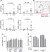
Macrophages in the ascites of epithelial ovarian carcinoma (EOC) and control patients. (a) Immunohistochemical staining of the cytospin specimens of advanced EOC ascites. Many CD68+ and CD163+ macrophages are observed. (b,c) The numbers of CD68+ and CD163+ macrophages in the ascites. (Kruskal–Wallis test) [B]: benign disease, [I]: EOC patients of Stage I, [III, IV]: Stages III and IV. n.s. not significant.
Figure 2
Figure 2
Proliferation of SKOV3 cells and human ovarian surface epithelial (hOSE) cells in the presence of ascitic fluids. The ascites of advanced epithelial ovarian carcinoma (EOC) induced strong proliferation of SKOV3 cells and hOSE cells. Cell numbers are expressed as the percentage of untreated controls. [B]: benign disease, [I]: EOC patients of Stage I, [III, IV]: Stages III and IV.
Figure 3
Figure 3
Cytokine levels in ascites and effect of cytokines on tumor proliferation. (a) Increased levels of interleukin (IL)‐6, IL‐10, growth‐related oncogene alpha (GRO‐α), and vascular endothelial growth factor (VEGF)‐A were detected in advanced epithelial ovarian carcinoma (EOC) ascites (Kruskal–Wallis test). [B]: benign disease, [I]: EOC patients of Stage I, [III, IV]: Stages III and IV. (b) Double immunostaining of cell‐block of ascites revealed that CD163+ macrophages as well as CD163 cancer cells produced IL‐6 in advanced EOC ascites. Scale bar = 50 μm. (c) SKOV3 cells were cultured with recombinant IL‐6, IL‐10, and GRO‐α, and the cell proliferation was evaluated by a WST assay. SKOV3 cells proliferated significantly in the presence of these cytokines. (d) Neutralizing antibodies against IL‐6 and IL‐10 were added with ascites, and SKOV3 cells were cultured for 5 days. Proliferation of SKOV3 cells was significantly suppressed by simultaneous blocking of IL‐6 and IL‐10. The results are expressed as the percentage of untreated controls. (Mann–Whitney test, *P < 0.01, **P < 0.05).
Figure 4
Figure 4
Stat3 activation and interleukin (IL)‐10 production in THP‐1 cells by ascitic fluids. (a, b) Following the culture of THP‐1 cells in the presence of 20% ascites for 30 min, Stat3 activation was evaluated immunohistochemically using anti‐phosphorylated Stat3 (pStat) antibody. Distinct staining for pStat3 was observed in the nuclei of THP‐1 cells treated with epithelial ovarian carcinoma (EOC) ascites. Cont: no ascites added. SKOV3: SKOV3 cell‐supernatant used as a positive control. (c) THP‐1 macrophages were cultured in the presence of 20% ascites for 2 days, then the cells were stimulated with lipopolysaccharide (100 ng/mL) for 24 h. IL‐10 production was analyzed by means of ELISA. A significant increase in the IL‐10 production was induced by the ascites of advanced EOC patients. [B]: benign disease, [I]: EOC patients of Stage I, [III, IV]: Stages III and IV. Statistical differences were evaluated by using Mann–Whitney test (b,c).
Figure 5
Figure 5
Stat3 activation by cell‐to‐cell interactions. (a) SKOV3 cells and primary monocyte‐derived macrophages were mixed and cultured for 5 days. Cells were detached and prepared as cell block specimens. Stat3 activation and cyclin‐D1, a molecule associated with cell proliferation, were analyzed via double immunostaining. (b) SKOV3 cells and monocyte‐derived macrophages were cultured separately. Little or no Stat3 activation was detected in both experiments. Scale bar = 50 μm. (c) Mixed culture of SKOV3 cells and macrophages (pretreated with macrophage colony stimulating factor [M‐CSF] or granulocyte‐macrophage colony stimulating factor [GM‐CSF] from monocytes). Stat3 activation was analyzed using double immunostaining of cell‐block specimens. Stat3 activation is seen in the nuclei of CD68+ macrophages and CD68 SKOV3 cells. The arrowheads indicate CD68+ macrophages with Stat3 activation. Scale bar = 50 μm. (n = 3 for each group) (d) Cyclin‐D1 expression in mixed culture of SKOV3 cells and macrophages (pretreated with M‐CSF or GM‐CSF from monocytes). Cyclin‐D1 was analyzed by double immunostaining. Many cyclin‐D1+ cells were found in CD68 SKOV3 cells by double immunostaining. Scale bar = 50 μm. The percentage of cyclin‐D1+ SKOV3 cells was higher in co‐culture than in single culture. (n = 3 for each group) (e) SKOV3 cells and macrophages were co‐cultured without actual cellular contact using a transwell co‐culture system. Stat3 activation was evaluated by a Western blot assay. Stat3 activation in both cell types was observed in the transwell co‐culture system (Mann–Whitney test, *P < 0.01, **P < 0.05).
Figure 6
Figure 6
SiRNA mediated knockdown of endogenous Stat3 in macrophages (a) Two days after suppression of Stat3 in macrophages, SKOV3 cells were added to the culture plate. After co‐culture for 5 days, cells were prepared as cell block specimens. (b) The Stat3 expression was evaluated by a Western blot analysis. Stat3 expression in macrophages differentiated with granulocyte‐macrophage colony stimulating (GM‐CSF) was suppressed by siRNA. (c) Two days after suppression of Stat3 in macrophages, cells were washed in PBS to deplete the siRNA. After co‐culture for 3 days with SKOV3 cells, cells were prepared as cell‐block specimens for immunostaining. Stat3 activation in SKOV3 was significantly suppressed by pretreatment of macrophages with siRNA. Scale bar = 50 nm. Similarly, Cyclin‐D1 expression in SKOV3 cells (CD68 cells) was also suppressed by siRNA pretreatment. (d) Simultaneous blocking of interleukin (IL)‐6 and IL‐10 by neutralizing antibodies induced significantly suppression of Stat3 activation in SKOV3 cells co‐cultured with macrophages. (e) Cultured human macrophages were stimulated by SKOV3 culture supernatant for 2 days, and the mRNA expressions of IL‐6, IL‐10, and CD163 were examined by quantitative PCR (Q‐PCR). The expression of these molecules was significantly suppressed by Stat3 siRNA. (Mann–Whitney test, *P < 0.01, **P < 0.05) (n = 3 for each group).
Figure 7
Figure 7
Involvement of Stat3 in growth of SKOV3 cells. (a) Western blot analysis shows that Stat3 expression in SKOV3 cells is suppressed by siRNA. (b) Following siRNA treatment, SKOV3 was cultured for 5 days and the viability of SKOV3 cells was evaluated by WST‐1 assay. The proliferation of SKOV3 was significantly suppressed by down‐regulation of Stat3 (n = 3 for each group, Mann–Whitney test, *P < 0.01). n.s. not significant.

References

    1. Okamura H, Katabuchi H. Pathophysiological dynamics of human ovarian surface epithelial cells in epithelial ovarian carcinogenesis. Int Rev Cytol 2005; 242: 1–54. - PubMed
    1. Bhoola S, Hoskins WJ. Diagnosis and management of epithelial ovarian cancer. Obstet Gynecol 2006; 107: 1399–1410. - PubMed
    1. Shield K, Ackland ML, Ahmed N, Rice GE. Multicellular spheroids in ovarian cancer metastases: biology and pathology. Gynecol Oncol 2009; 113: 143–148. - PubMed
    1. Okamura H, Katabuchi H, Kanzaki H. Macrophages in reproductive biology. In: Lewis C, Burke B, eds. The Macrophages. London: Oxford University Press, 2002; 548–576.
    1. Wang X, Deavers M, Patenia R et al. Monocyte/macrophage and T‐cell infiltrates in peritoneum of patients with ovarian cancer or benign pelvic disease. J Transl Med 2006; 4: 30. - PMC - PubMed

Publication types