. 2010 Sep 22:11:37.
doi: 10.1186/1471-2091-11-37.

The effects of oligomerization on Saccharomyces cerevisiae Mcm4/6/7 function

Affiliations

The effects of oligomerization on Saccharomyces cerevisiae Mcm4/6/7 function

Xiaoli Ma et al. BMC Biochem. .

Abstract

Background: Minichromosome maintenance proteins (Mcm) 2, 3, 4, 5, 6 and 7 are related by sequence and form a variety of complexes that unwind DNA, including Mcm4/6/7. A Mcm4/6/7 trimer forms one half of the Mcm2-7 hexameric ring and can be thought of as the catalytic core of Mcm2-7, the replicative helicase in eukaryotic cells. Oligomeric analysis of Mcm4/6/7 suggests that it forms a hexamer containing two Mcm4/6/7 trimers, however, under certain conditions trimeric Mcm4/6/7 has also been observed. The functional significance of the different Mcm4/6/7 oligomeric states has not been assessed. The results of such an assessment would have implications for studies of both Mcm4/6/7 and Mcm2-7.

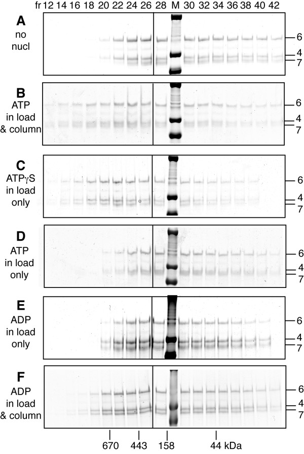
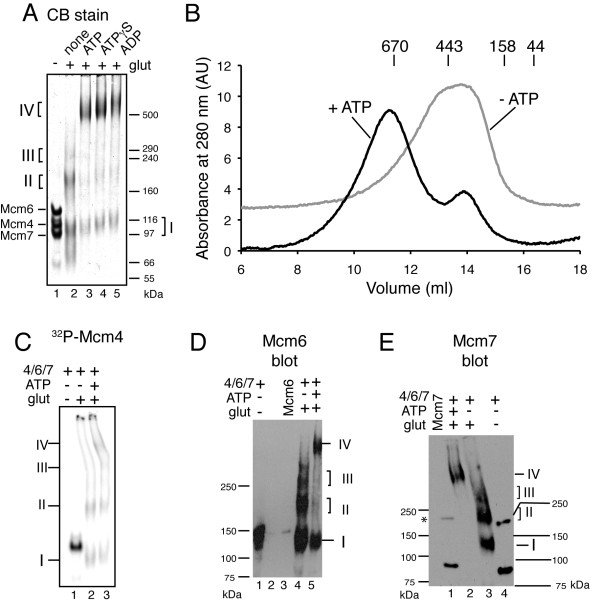
Results: Here, we show that Saccharomyces cerevisiae Mcm4/6/7 reconstituted from individual subunits exists in an equilibrium of oligomeric forms in which smaller oligomers predominate in the absence of ATP. In addition, we found that ATP, which is required for Mcm4/6/7 activity, shifts the equilibrium towards larger oligomers, likely hexamers of Mcm4/6/7. ATPγS and to a lesser extent ADP also shift the equilibrium towards hexamers. Study of Mcm4/6/7 complexes containing mutations that interfere with the formation of inter-subunit ATP sites (arginine finger mutants) indicates that full activity of Mcm4/6/7 requires all of its ATP sites, which are formed in a hexamer and not a trimer. In keeping with this observation, Mcm4/6/7 binds DNA as a hexamer.

Conclusions: The minimal functional unit of Mcm4/6/7 is a hexamer. One of the roles of ATP binding by Mcm4/6/7 may be to stabilize formation of hexamers.


Figures

Figure 1
Figure 1
Reconstitution of Mcm4/6/7 using anion exchange chromatography. A) A portion of the indicated fractions from the Mono Q column were analyzed by SDS-PAGE (6%) and stained with Coomassie Brilliant Blue. The migration of size markers through the gel is indicated on the left. The migrations of Mcm4, Mcm6 and Mcm7 are indicated on the right. There is a minor contaminating band that migrates between Mcm6 and Mcm4 that is found in all of our preparations. B) ATP hydrolysis (black square) and DNA unwinding (white circle) by equal volumes of the indicated fractions were analyzed as described in the "Methods" section. C) A 50 μg portion of Mono Q fraction 67 was analyzed by gel filtration on a Superose 6 PC 3.2/30 column equilibrated in Buffer A with 100 mM NaCl. Shown here are Colloidal Coomassie Blue-stained SDS polyacrylamide gels (6%) of the indicated fractions. The vertical line marks the border between separate gels. The peak elutions of size standards are indicated at the bottom of the gel. The migration of size standards though the SDS gels is indicated on the left and of Mcm4, Mcm6 and Mcm7 on the right.
Figure 2
Figure 2
ATP hydrolysis by individual and pairwise Mcms and Mcm4/6/7 complexes. The mean rate of ATP hydrolysis for the indicated individual Mcms, pairwise combinations and Mcm4/6/7 complexes is shown. Rate measurements were determined at 1 mM ATP as described in "Methods". For the pairwise combinations of Mcm proteins, the rate is expressed per μg of the Mcm protein that is predicted to bind nucleotide within the pairs (as indicated). For comparison to Mcm4 + Mcm7, the rate of ATP hydrolysis by Mcm4/6/7 is expressed per μg Mcm4 (assuming equimolar ratios of each protein in the complex).
Figure 3
Figure 3
Isolation of reconstituted Mcm4/6/7 by size exclusion chromatography. A) A typical elution profile of Mcm4/6/7 from a Superose 6 10/300 GL column. A portion of the indicated fractions was analyzed by Coomassie Brilliant Blue-stained SDS-PAGE (6%). The vertical line marks the border between separate gels. The peak elution of size standards is indicated. The migration of size standards through the SDS gels is indicated on the left and of Mcm4, Mcm6 and Mcm7 on the right. B) Equal volumes of the indicated fractions were analyzed for ATP hydrolysis (white square) or DNA unwinding (black circle) as described in "Methods". C) The absorbance at 280 nm (A280) corresponding to the elution of each of Mcm4, Mcm6 and Mcm7 from the gel filtration column are shown. The peaks in absorbance correspond to the peak in the indicated proteins as determined by SDS-PAGE (not shown). The A280 curves of size standards (apoferritin, 443 kDa; BioRad standards which include thyroglobulin, 670 kDa; bovine gamma-globulin, 158 kDa and chicken ovalbumin, 44 kDa) are also shown.
Figure 4
Figure 4
Effect of different nucleotides on oligomerization of Mcm4/6/7. The elution of Mcm4/6/7 from a 2.4 ml "mini" gel filtration column (Superose 6 PC 3.2/30; GE Healthcare) in the presence and absence of 5 mM adenine nucleotides is shown. A portion of the indicated fractions (above top panel) from each experiment was analyzed by GelCode Blue (Pierce) stained SDS-PAGE (6%). The migrations of Mcm4, Mcm6 and Mcm7 through the SDS gels are indicated to the right of each panel. Lanes containing molecular size markers are indicated (M) and their sizes are indicated on the left. The peak elution fractions of size standards (analyzed separately) are indicated below the bottom panel. The vertical lines mark the divisions between gels. In some experiments, nucleotide was added only to the protein (indicated by "load") and in some experiments nucleotide was added to both the protein and to the buffer used to equilibrate the gel filtration column ("load & column"). A) No nucleotide in either column buffer or load; B) 5 mM ATP in column buffer and load; C) 5 mM ATPγS added to load only; D) 5 mM ATP added to load only; E) 5 mM ADP in load only; and F) 5 mM ADP in column buffer and load. UV profiles are not shown since addition of nucleotide interfered with the signal from protein.
Figure 5
Figure 5
Chemical crosslinking of Mcm4/6/7. Crosslinking of Mcm4/6/7 with glutaraldehyde in the presence and absence of ATP, ADP or ATPγS is shown. A) The samples were analyzed on 3.5% SDS-phosphate gels stained with Coomassie Brilliant Blue ("CB stain"). Each lane contains 9 μg of Mcm4/6/7 in the presence and absence of glutaraldehyde and/or 1 mM ATP, ADP or ATPγS (as indicated). The migration of size standards is indicated on the right and of Mcm4, Mcm6 and Mcm7 in the absence of crosslinker is indicated on the left as are the migration of crosslinked products (by Roman numerals). B) Mcm4/6/7 was crosslinked with glutaraldehyde with and without 5 mM ATP before analysis on a Superose 6 10/300 GL column in the absence of added nucleotide. The A280 curves from the columns are shown for Mcm4/6/7 crosslinked in the presence of ATP (black line) and in the absence of ATP (grey line). The peak elution volumes of size standards, analyzed separately are shown above the graph. C) Phosphorimager scan of a SDS-phosphate gel of 32P-labeled Mcm4PK/6/7 crosslinked in the presence and absence of ATP (as indicated) is shown. The migrations of crosslinked products, based on Coomassie Blue staining of the same gel are indicated. Western blots to detect Mcm6 (D) and Mcm7 (E) in the crosslinked products. The migrations of Mcm6 and Mcm7 in the absence of crosslinker as well as the migration of crosslinked products are indicated. The presence of glutaraldehyde and ATP are indicated at the top of the blots. Standards with Mcm6 alone (40 ng) or Mcm7 alone (20 ng) are also indicated. The asterisk indicates a cross-reacting band with Mcm7 antibody that is barely visible in the Coomassie stained gels (see A).
Figure 6
Figure 6
Isolation and activity of Mcm4/6/7 complexes containing mutant proteins. A) Models for the subunit arrangement in a Mcm4/6/7 hexamer (left) and trimer (right) are depicted. The complexes are drawn viewing the C-terminal face of the ring and are based on known physical interactions between S. cerevisiae MCM proteins (modified from [35]). "SRF" indicates the conserved motif encoding the arginine finger. Other arrangements of the Mcm4/6/7 hexamer are formally possible, however these alternatives are less likely than the model shown given the current knowledge of Mcm and related protein structures and the known physical interactions between Mcm proteins [10,14,35,47,61]. B) Mcm4/6/7 complexes were reconstituted using all wild-type proteins ('WT'), Mcm4R701A, Mcm6 and Mcm7 ("4RA/6/7"); Mcm4, Mcm6R703A and Mcm7 ("4/6RA/7"); and Mcm4, Mcm6 and Mcm7R593A ("4/6/7RA") and the complexes were isolated by size exclusion chromatography as described in "Methods". The "WT" experiment shown here for comparison is from Figure 2. A portion of the indicated fractions was visualized by Coomassie Brilliant Blue stained SDS-PAGE (6%) to determine the peak fractions. The migrations of Mcm4, Mcm6 and Mcm7 (mutant or wild-type) through the gels are indicated to the right of each panel. The peak elution fractions of size standards analyzed separately are indicated below the bottom panel. C) DNA unwinding by wild-type and mutant Mcm4/6/7 complexes was compared. Each assay contained 750 ng of Mcm4/6/7. The numbers above the bars in the graph correspond to the ATP sites in part A. D) ATP hydrolysis was determined as described in "Methods". The mean rate per μg of Mcm4/6/7 is shown. The numbers above the bars in the graph correspond to the ATP sites shown in the hexamer model in part A and indicate which site is affected by the mutation.
Figure 7
Figure 7
Mcm4/6/7 binds single stranded DNA as a hexamer. Mcm4/6/7 was crosslinked in the presence of single stranded DNA using glutaraldehyde. The indicated amounts of Mcm4/6/7 were incubated with 20 nM 32P-dT60 and 1 mM ATP in 6 μl of 20 mM HEPES NaOH pH 7.5 and 10 mM magnesium acetate. After incubation at 25°C for 10 min, 0.4 μl of 2.3% glutaraldehyde was added. The mixture was then incubated at 37°C for 2 min before quenching the reaction with 0.8 μl 2 M Tris. After addition of 4 μl 60% glycerol, the samples were analyzed on a 5% polyacrylamide gel in 0.5 × TBE. A) A phosphorimager scan of crosslinking of Mcm4/6/7 to dT60 in the presence of ATP. The migration of size markers, based on Coomassie Brilliant Blue staining of the gel is indicated on the right. Note that in order to resolve trimer and hexamer sized, the gel was run so that free DNA migrated off the bottom of the gel. B) Crosslinking of Mcm4/6/7 in the presence and absence of DNA (as indicated). A Phosphorimage is shown on the right and the same gel, stained with GelCode Blue (Pierce) is on the left. Migration of size standards is shown on the right. C). Crosslinking of Mcm4/6/7 in the presence and absence of 1 mM ATP, as indicated. Migration of size standards is shown on the right. Lane 3 is an empty lane.

Similar articles

Cited by

References

    1. Singleton MR, Dillingham MS, Wigley DB. Structure and mechanism of helicases and nucleic acid translocases. Annual Review of Biochemistry. 2007;76:23–50. doi: 10.1146/annurev.biochem.76.052305.115300. - DOI - PubMed
    1. Kaplan DL, Davey MJ, O'Donnell M. Mcm4,6,7 uses a "pump in ring" mechanism to unwind DNA by steric exclusion and actively translocate along a duplex. J Biol Chem. 2003;278:49171–49182. doi: 10.1074/jbc.M308074200. - DOI - PubMed
    1. Laskey RA, Madine MA. A rotary pumping model for helicase function of MCM proteins at a distance from replication forks. EMBO Rep. 2003;4:26–30. doi: 10.1038/sj.embor.embor706. - DOI - PMC - PubMed
    1. Lee JK, Hurwitz J. Processive DNA helicase activity of the minichromosome maintenance proteins 4, 6, and 7 complex requires forked DNA structures. Proc Natl Acad Sci USA. 2001;98:54–59. doi: 10.1073/pnas.98.1.54. - DOI - PMC - PubMed
    1. Takahashi TS, Wigley DB, Walter JC. Pumps, paradoxes and ploughshares: mechanism of the MCM2-7 DNA helicase. Trends in Biochemical Sciences. 2005;30:437–444. doi: 10.1016/j.tibs.2005.06.007. - DOI - PubMed

Publication types

MeSH terms

Substances

LinkOut - more resources