. 2010 Sep 22:7:15.
doi: 10.1186/1743-8454-7-15.

Aquaporins: relevance to cerebrospinal fluid physiology and therapeutic potential in hydrocephalus

Affiliations

Aquaporins: relevance to cerebrospinal fluid physiology and therapeutic potential in hydrocephalus

Brian K Owler et al. Cerebrospinal Fluid Res. .

Abstract

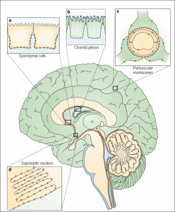
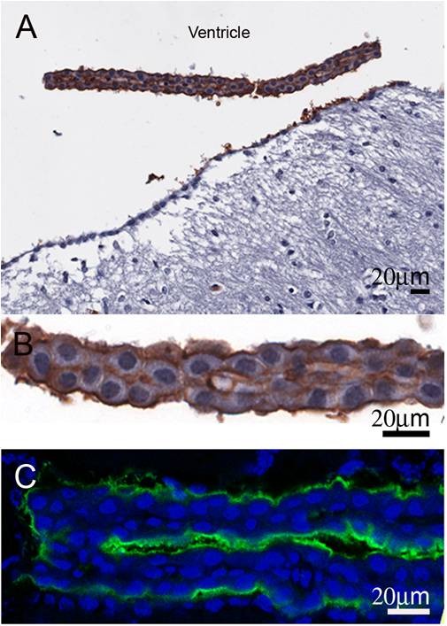
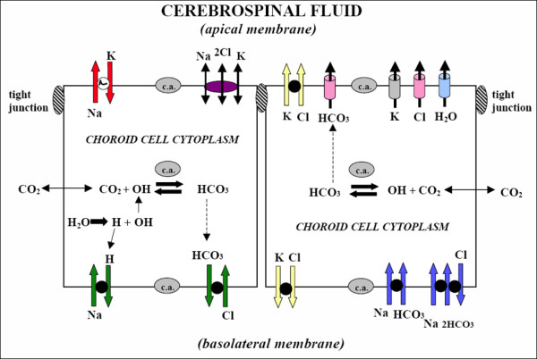
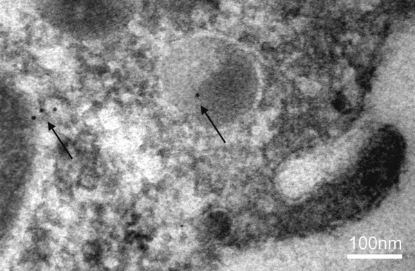
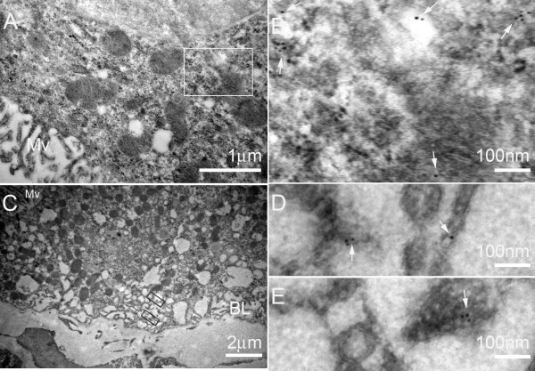
The discovery of a family of membrane water channel proteins called aquaporins, and the finding that aquaporin 1 was located in the choroid plexus, has prompted interest in the role of aquaporins in cerebrospinal fluid (CSF) production and consequently hydrocephalus. While the role of aquaporin 1 in choroidal CSF production has been demonstrated, the relevance of aquaporin 1 to the pathophysiology of hydrocephalus remains debated. This has been further hampered by the lack of a non-toxic specific pharmacological blocking agent for aquaporin 1. In recent times aquaporin 4, the most abundant aquaporin within the brain itself, which has also been shown to have a role in brain water physiology and relevance to brain oedema in trauma and tumours, has become an alternative focus of attention for hydrocephalus research. This review summarises current knowledge and concepts in relation to aquaporins, specifically aquaporin 1 and 4, and hydrocephalus. It also examines the relevance of aquaporins as potential therapeutic targets in hydrocephalus and other CSF circulation disorders.


Figures

Figure 1
Figure 1
Distribution in brain of aquaporin-1 (AQP1, blue) and AQP4 (orange), schematically illustrated on a sagittal section of a human brain. a: AQP4 occurs in the basolateral membrane of ependymal cells. b: AQP1 is expressed at the apical membrane of choroid plexus epithelial cells. c: AQP4 is concentrated in astrocytic end-feet, specifically in those membrane domains that abut on brain capillaries or on pia. d: AQP4 is expressed in glial lamellae of the supraoptic nucleus and other osmosensitive regions. AQP4 also occurs in non end-feet membranes of astrocytes, but at comparatively low concentrations. In the neocortex, AQP4 expression in non end-feet membranes increases from deep to superficial layers. The cerebellum shows the opposite gradient, with higher concentrations in the granule cell layer than in the molecular layer. Reprinted with permission from [20].
Figure 2
Figure 2
AQP1 localisation in choroid plexus epithelial cell in wild-type mice. Mouse coronal paraffin sections were incubated with rabbit anti-AQP1 antibody at 4°C overnight followed by incubation of anti-rabbit Ig conjugated with biotin and standard ABC technique (A and B) or anti-rabbit Ig conjugated with Alexa 488 (C). Positive staining for AQP1 is on the apical surface of the epithelial cells of normal wild-type mice. Panel B is the high magnification image of panel A. (Owler et al, unpublished data)
Figure 3
Figure 3
Ion transporters and channels in mammalian choroidal epithelium. CSF secretion results from coordinated transport of ions and water from basolateral membrane to cytoplasm, then sequentially across apical membrane into ventricles. On the plasma-facing membrane is parallel Na+-H+ and Cl--HCO3- exchange bringing Na+ and Cl- into cells in exchange for H+ and HCO3-, respectively. Also basolaterally located is Na+-HCO3- cotransport (NBCn1) and Na-dependent Cl--HCO3- exchange that modulate pH and perhaps CSF formation. Apical Na+ pumping maintains a low cell Na+ that sets up a favorable basolateral gradient to drive Na+ uptake. Na+ is extruded into CSF mainly via the Na+ pump and, under some conditions, the Na+-K+-2Cl- cotransporter. K+-Cl- cotransport helps maintain cell volume. Apical channels facilitate K+, Cl- and HCO3- diffusion into CSF. Aquaporin 1 (AQP1) channels on CSF-facing membrane mediate water flux into ventricles. Polarized distribution of carbonic anhydrase (c.a.) and Na+-K+-ATPase, and aquaporins, enable net ion and water translocation to CSF. Reprinted with permission from [14].
Figure 4
Figure 4
Immuno-gold electron micrograph of choroid plexus epithelial cell from a wild type mouse after cisternal kaolin injection. There is gold labelling (arrows) of AQP1 within the cytoplasm and a lysosome. (Owler et al, unpublished data).
Figure 5
Figure 5
Representative immuno-electron micrographs of choroidal epithelium in kaolin-injected hydrocephalic WT mice. A and C: low magnification image of choroidal epithelium of hydrocephalic mice. B, D and E: Higher magnification electron micrographs corresponding to the boxes in A and C show gold particles (white arrows) in the cytoplasm (B) and in the basal membrane of the epithelia (D and E). Mv: microvilli; BL: basolateral membrane. (Owler et al, unpublished data).
Figure 6
Figure 6
Histograms representing mean ventricular areas of the wild-type (WT)(Blue) and AQP1-null mice (AQP1 KO)(Red) at 3 and 5 days post kaolin injection (dpi) compared to saline injected control (con) mice. AQP1-null control mice (n = 14) had lower ventricular areas compared to wild-type control mice (n = 11). AQP1-null mice (n = 6 at 3 dpi; n = 11 at 5 dpi) did not demonstrate the same degree of ventricular dilation as wild-type mice (n = 8 at 3 dpi; n = 16 at 5 dpi) at either 3 or 5 days after cisternal kaolin injection. Data are means +/- SEM. (Owler et al, unpublished data).
Figure 7
Figure 7
Aquaporin-4 protein expression in mouse model of hydrocephalus. Aquaporin-4 immunostaining of AQP4+/+ control mouse (A), AQP4+/+ (B), and AQP4-/- (C) mice 5 days after kaolin injection. Asterisks indicate lateral ventricle, white arrowheads indicate ventricular ependyma. (D) Immunoblot of AQP4 protein expression in AQP4+/+ and AQP4-/- control mice and AQP4+/+ mice at 5 days after kaolin injection. Scale bar = 100 μm. Reprinted with permission from [60].
Figure 8
Figure 8
Lentiviral-GFP localisation in choroid plexus epithelial cells in mice. Normal Quackenbush Swiss mice received intraventricular injection of lentiviral-GFP (107 virus genomes in 10 μL of sterile saline) and harvested after 7 days of injection. Frozen coronal section of the brain were incubated with rabbit GFP antibody at 4°C overnight followed by incubation of anti-rabbit Ig conjugated with Alexa 488. GFP signals were found in the cytoplasm of some choroid epithelial cells (arrows) in lateral choroid plexus in panel A (confocal image) and C (composite of A and B). Panel B is the phase contrast image of the same field in A. (Owler et al., unpublished data).

References

    1. King LS, Agre P. Pathophysiology of the aquaporin water channels. Ann Rev Physiol. 1996;58:619–648. doi: 10.1146/annurev.ph.58.030196.003155. - DOI - PubMed
    1. Saboori AM, Smith BL, Agre P. Polymorphism in the Mr 32,000 Rh protein purified from Rh(D)-positive and -negative erythrocytes. Proc Natl Acad Sci USA. 1988;85:4042–4045. doi: 10.1073/pnas.85.11.4042. - DOI - PMC - PubMed
    1. Preston GM, Carroll TP, Guggino WB, Agre P. Appearance of water channels in Xenopus oocytes expressing red cell CHIP28 protein. Science. 1992;256:385–387. doi: 10.1126/science.256.5055.385. - DOI - PubMed
    1. Nielsen S, Smith BL, Christensen EI, Agre P. Distribution of the aquaporin CHIP in secretory and resorptive epithelia and capillary endothelia. Proc Natl Acad Sci USA. 1993;90:7275–7279. doi: 10.1073/pnas.90.15.7275. - DOI - PMC - PubMed
    1. Praetorius J, Nielsen S. Distribution of sodium transporters and aquaporin-1 in the human choroid plexus. Am J Physiol Cell Physiol. 2006;291:C59–67. doi: 10.1152/ajpcell.00433.2005. - DOI - PubMed

LinkOut - more resources