. 2010 Sep 22:6:60.
doi: 10.1186/1744-8069-6-60.

Endogenous N-acetylaspartylglutamate (NAAG) inhibits synaptic plasticity/transmission in the amygdala in a mouse inflammatory pain model

Affiliations

Endogenous N-acetylaspartylglutamate (NAAG) inhibits synaptic plasticity/transmission in the amygdala in a mouse inflammatory pain model

Mary O Adedoyin et al. Mol Pain. .

Abstract

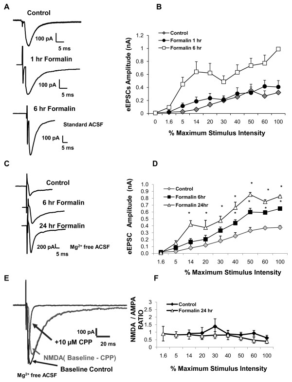
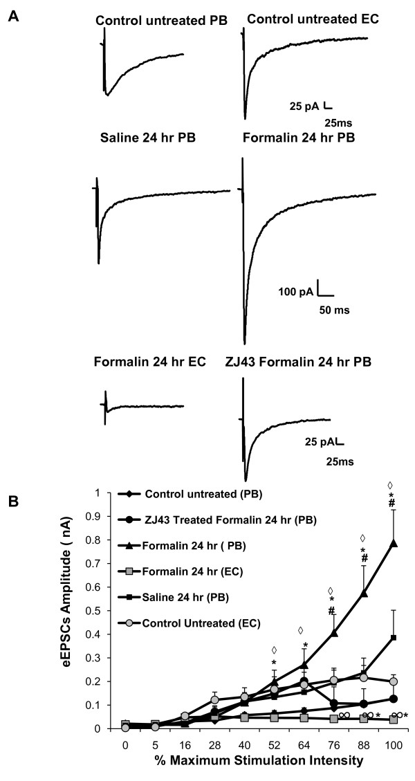
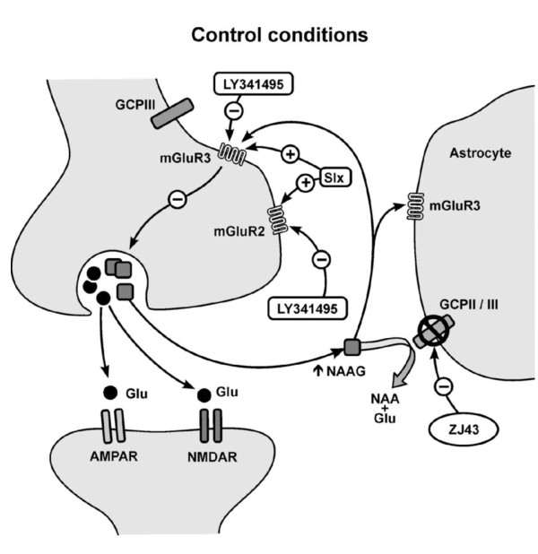
Background: The peptide neurotransmitter N-acetylaspartylglutamate (NAAG) is widely expressed throughout the vertebrate nervous system, including the pain processing neuraxis. Inhibitors of NAAG peptidases are analgesic in animal models of pain. However, the brain regions involved in NAAG's analgesic action have not been rigorously defined. Group II metabotropic glutamate receptors (mGluR2/3) play a role in pain processing in the laterocapsular part of the central nucleus of the amygdala (CeLC). Given the high concentration of NAAG in the amygdala and its activation of group II mGluRs (mGluR3 > mGluR2), this study was undertaken using the mouse formalin model of inflammatory pain to test the hypothesis that NAAG influences pain processing in the amygdala. Evoked excitatory postsynaptic currents (eEPSCs) were studied in neurons in the CeLC of mouse brain slices following stimulation of the spinoparabrachial amygdaloid afferents.

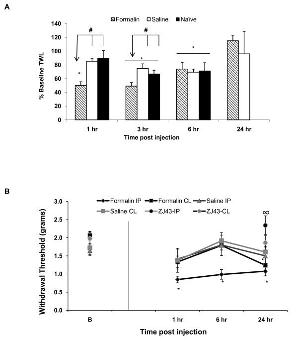
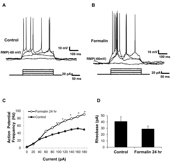
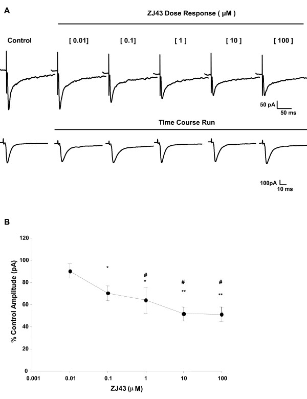
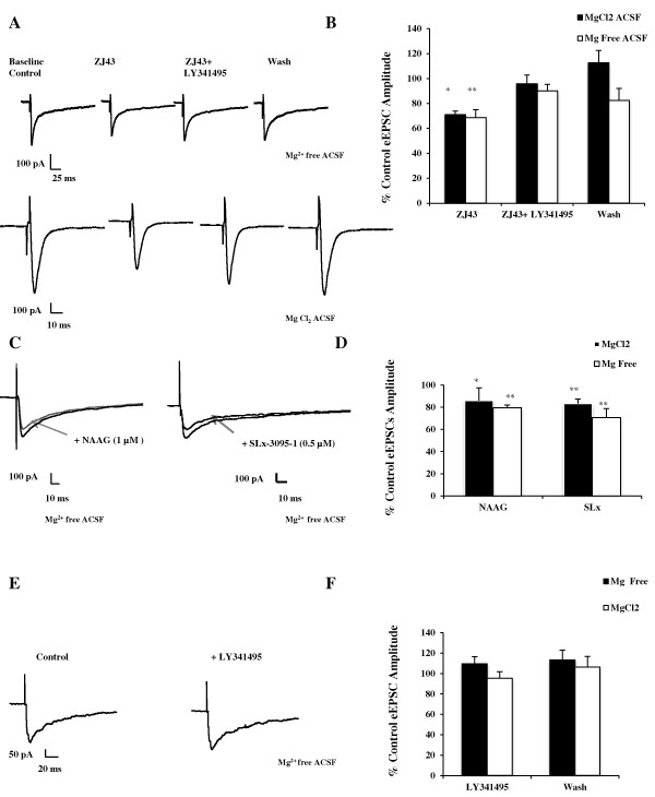
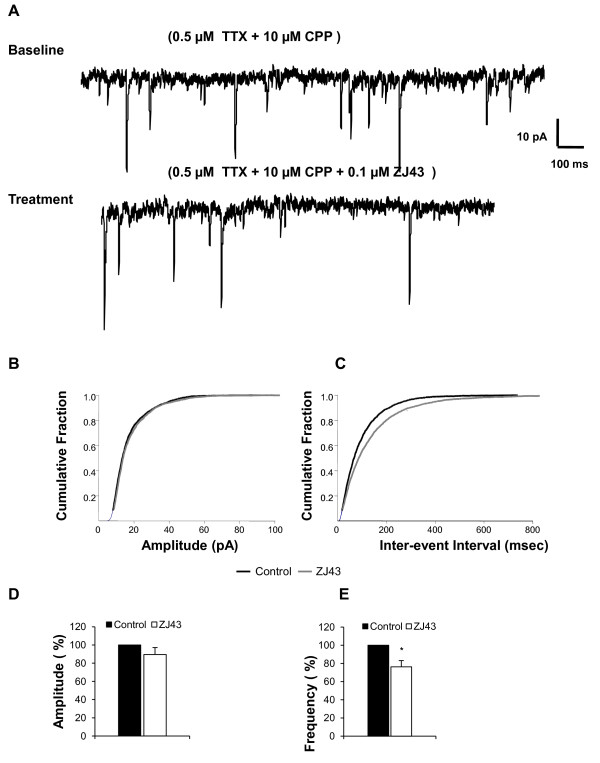
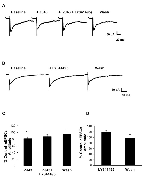
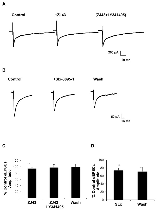
Results: Application of a NAAG peptidase inhibitor, ZJ43, dose dependently inhibited the amplitude of the eEPSCs by up to 50% in control CeLC demonstrating the role of NAAG in regulation of excitatory transmission at this synapse. A group II mGluR agonist (SLx-3095-1) similarly inhibited eEPSC amplitude by about 30%. Both effects were blocked by the group II mGluR antagonist LY341495. ZJ43 was much less effective than SLx in reducing eEPSCs 24 hours post inflammation suggesting an inflammation induced reduction in NAAG release or an increase in the ratio of mGluR2 to mGluR3 expression. Systemic injection of ZJ43 proximal to the time of inflammation blocked peripheral inflammation-induced increases in synaptic transmission of this pathway 24 hrs later and blocked the induction of mechanical allodynia that developed by this time point.

Conclusions: The main finding of this study is that NAAG and NAAG peptidase inhibition reduce excitatory neurotransmission and inflammation-induced plasticity at the spinoparabrachial synapse within the pain processing pathway of the central amygdaloid nucleus.


Figures

Figure 1
Figure 1
Prolonged nociceptive behaviors in formalin mice model. (A) Baseline thermal withdrawal latency (TWL) was determined for each mouse prior to treatment. This value was used to establish 100% baseline for that subject. Control groups (saline and naïve) mice showed habituation when retested at 3 and 6 hours. Thermal hypersensitivity (lower TWL relative to baseline as 100%) is observed in the formalin treated mice (n = 15) compared to the control groups (saline-treated n = 11; naïve n = 10) at 1 and 3 but not 6 hours post injection. The formalin treated group TWL was significantly different at 1 hr compared to naïve (p < 0.001) and (p < 0.005) saline groups; at 3 hr compared to saline (p < 0.02) and naïve (p < 0.05) groups. TWL returns to baseline values at 24 hours in the treated groups. #p ≤ 0.05 across group comparison for TWL values by two way RM-ANOVA followed by pairwise Tukey t-test post hoc analysis. (B) Mechanical allodynia, measures as paw withdrawal in response to force of probe in grams, was assessed in the injected hindpaws (ipsilateral = injected paw, IP) and opposite (contralateral, CL) hindpaws. There was a significant difference between the ipsilateral and contralateral footpad (p < 0.02) in the formalin mice. Ipsilateral hindpaw allodynia was significant at 1, 6 and 24 hours post-inflammation relative to the baseline values (n = 15). At 24 hours post-inflammation, the contralateral threshold was significantly (p < 0.05) lower compared to the baseline values in the contralateral footpad. ZJ43 (150 mg/kg; IP, n = 10) administered 30 prior to and 8 hours post formalin injection significantly reduced allodynia in the ipsilateral footpad (∞p ≤ 0.05, ZJ43 (formalin)-IP vs. Formalin-IP mechanical withdrawal threshold values at 24 hr post inflammation). The difference between ZJ43 (formalin)-CL and Formalin-CL did not reach statistical significance (p = 0.13). Two way RM-ANOVA was used followed by pairwise Tukey t-test post hoc analysis. Baseline (B) = probe force that induced paw withdrawal in mice prior to injection of formalin or saline. Data presented as mean ± SEM, *p ≤ 0.05 vs. baseline values; was used followed by pairwise Tukey t-test post hoc analysis.
Figure 2
Figure 2
Enhanced synaptic transmission in the CeLC nucleus of control and formalin brain slices. Enhanced evoked current was observed in the central amygdala, laterocapsular part (CeLC) neurons 6 and 24 hours after peripheral inflammation. Input-output experiments were conducted to examine neurotransmission changes in the CeLC as a result of peripheral inflammation (QX-314 [5 mM] local anesthetic was included in the pipette solution). (A) Representative trace depicting evoked response at maximal stimulation intensity in cells from untreated control and formalin treated animals 1 and 6 hours post inflammation (standard ACSF).(B) An increase in current output was observed at 6 hours (n = 6) and not 1 hour (n = 9-13) in the CeLC post inflammation.(C) Representative trace depicting evoked response at maximal stimulation intensity in cells from untreated control and formalin treated animals 6 and 24 hours post inflammation (Mg2+ Free ACSF). (D) There was a significant gain in the current output at 6 (n = 13) and 24 hours (n = 12) in the CeLC following inflammation. (E) Representative trace depicts how NMDA/AMPA ratio was obtained. NMDAR antagonist, CPP (10 μM), was used to determine the relative contribution of the NMDA and AMPA components of the evoked current response during input-output experiments (Mg2+ Free ACSF). (F) NMDA/AMPA ratio was unchanged in the CeLC 24 hours after inflammation [control (n = 6), formalin 24 hours (n = 11)]. Data presented as mean ± SEM, *p ≤ 0.05 vs. control, Student's t-test.
Figure 3
Figure 3
Enhanced neuronal excitability in the CeLC neurons in brain slice of formalin-treated mice. CeLC neurons in slices from untreated control and formalin treated mice were tested 24 hours after inflammation by injection with 20 pA current steps. Example of CeLC neuron firing patterns of untreated control (A), and formalin-treated mice (B) in response to the depolarizing current steps. (C) Average data showing increased action potential frequency in the CeLC neurons in brain slices 24 hours after inflammatory insult compared to untreated control [Formalin (n = 18), Control (n = 30)].(D) Average rheobase current in CeLC neurons in brain slices [Control (n = 28), Formalin (18)]. Data presented as mean ± SEM, *p ≤ 0.05 vs. control, Student's t-test. Recordings were in Mg2+ free ACSF.
Figure 4
Figure 4
Dose response effect of ZJ43, peptidase inhibitor, on evoked current in control mouse CeLC region. (A) Representative evoked current traces shown in upper panel of ZJ43 at each concentration in a CeLC neuron; lower panel shows a representative cell current output traces over the same time duration as dose response experiments without a significant rundown in magnitude. (B) Average values for ZJ43 dose response effect on evoked excitatory currents in the CeLC of the untreated control mouse brain slice (n = 4). ZJ43 (10 μM) resulted in maximal (~50%) reduction in the amplitude of the evoked current. ZJ43's inhibitory effect at 100 μM (p < 0.01), 10 μM (p < 0.02) and 1 μM (p < 0.01) were significantly more potent compared to its effect at the minimum dose 10 nM. Data presented as mean ± SEM of percent control values, *p ≤ 0.05 vs. control, #p ≤ 0.05 vs. 10 nM ZJ43, one way RM-ANOVA followed by pairwise Tukey's t-test post hoc analysis. Recordings were in Mg2+ free ACSF.
Figure 5
Figure 5
Endogenous and applied NAAG inhibit synaptic transmission in the CeLC of the mouse amygdala. (A) Representative current traces recorded from two neurons in the CeLC region of brain slices from untreated control mice following stimulation of the spinoparabrachial amygdaloid afferents in the absence and presence of MgCl2 in the ACSF. ZJ43 (0.1 μM) inhibits inactivation of endogenously released NAAG and significantly reduces the amplitude of the evoked excitatory current. This effect is reversed by the group II mGluR antagonist LY341495 (1 μM). (B) Bar graph showing average data in each recording condition (n = 7, Mg2+ free ACSF; n = 6, standard ACSF). (C) Representative current traces from cells treated with NAAG (1 μM, n = 4) or group II mGluR agonist SLx-3095-1 (0.5 μM, n = 7). Both agonists inhibit the evoked current in the CeLC. (D) Bar graph showing average values from each drug treatment in the recording conditions. (E and F) Group II antagonist does not significantly affect evoked eEPSCs in control CeLC presented as mean ± SEM of percent control values. *p ≤ 0.05 vs. control, paired Student's t-test.
Figure 6
Figure 6
Endogenous NAAG acts presynaptically to reduce the frequency of mEPSCs in the CeLC region. Application of ZJ43 (0.1 μM) in the presence of TTX and CPP (to block action potentials and NMDA current respectively) in the CeLC region of the amygdala in slices from untreated control mice significantly reduced the frequency of miniature EPSC, while the amplitude of the mEPSCs was not significantly altered. A) Representative current traces in the absence and presence of ZJ43 in a neuron. (B) Cumulative amplitude curve in the presence and absence of ZJ43. (C) Cumulative inter-event interval curve depicting mEPSCs frequency rate in the presence and absence of ZJ43 in a neuron from a control untreated mouse brain slice. Bar graph showing % control of amplitude (p = 0.3, n = 9, ZJ43 treated versus control) (D) and frequency (E) values for cells (n = 9). Data presented as mean ± SEM, *p ≤ 0.05 vs. control, paired Student's t-test. Recordings were in Mg2+ free ACSF.
Figure 7
Figure 7
ZJ43 inhibit the evoked current in CeLC neurons 6 hours after induction of peripheral inflammation. The group II mGluR antagonist, LY341495 (1 μM), was used to define the level of tonic activation of the receptors as a consequence of peripheral inflammation. Representative traces: (A) ZJ43 (0.1 μM) significantly reduced eEPSCs in CeLC neurons; Average data showing (D) ZJ43 effects 6 hours post inflammation (n = 7). (B) In a separate set of cells, the direct effect of LY341495 was tested on the evoked current in the brain slice 6 hours post formalin treatment; (C) Average data showing LY341495 effect 6 hours post inflammation (p = 0.17, n = 7). Data presented as mean ± SEM percent control; *p ≤ 0.05 vs. control, paired Student's t-test. Recordings were in Mg2+ free ACSF.
Figure 8
Figure 8
The efficacy of ZJ43 and SLx-3095-1 in CeLC 24 hours post peripheral inflammation. Whole cell recordings were conducted to investigate the inhibitory efficacy of ZJ43 (0.1 μM) and SLx-3095-1 (0.5 μM) in the CeLC 24 hours post peripheral inflammation. (A) Representative trace of the ZJ43 effect. (C) Average data for experiments shown in A in which ZJ43 alone had very little effect on eEPSCs (5% reduction, p < 0.01, n = 10, paired Student's t-test in contrast to its effect in controls (Figure 4). (B) SLx-3095-1 significantly reduced the amplitude of the eEPSCs in the CeLC region (n = 12), average data shown in (D). Data presented as mean ± SEM percent control; *p ≤ 0.05 vs. control, paired Student's t-test. Recordings were in Mg2+ free ACSF.
Figure 9
Figure 9
PB-CeA and not EC-CeA synapse is activated in response to peripheral inflammation. To investigate the specificity of the PB-CeA pathway output to the inflammation, the external capsule (EC) was stimulated while recordings were made from neurons present in the CeLC region of brain slices obtained from formalin treated animals. For these stimulus intensity experiments, the local anesthetic QX-314 (5 mM) was included in the pipette solution. (A) The enhanced current output induced by inflammation observed after stimulation of the PB afferents (middle panel, n = 5) did not occur when the EC was stimulated (lower panel, n = 5). Stimulation of the EC in the formalin brain slices yielded significantly lower current magnitude at the higher intensity compared to that obtained in untreated control brain slices (upper panel, n = 4). Injection (i.p.) of NAAG peptidase inhibitor ZJ43(150 mg/kg, n = 6, lower panel) 30 minutes before and 8 hours after formalin injection into the mouse's footpad significantly blocked the enhanced current output that would have occurred with stimulation of the PB afferents at the higher stimulus intensities in the formalin group (middle panel). Larger current output albeit not significant was observed in the CeLC region of brain slices obtained from the saline group (middle panel, n = 5) when compared to the untreated control group (upper panel, n = 5). (B) Average data showing the current output as a function of the stimulus intensity in all four animal groups (untreated control, saline treated, ZJ43- formalin treated, and formalin treated). For the treated groups the recordings were performed 24 hours post saline or formalin injection into the mouse's footpad. Afferents stimulation: (PB) indicates the parabrachial afferents, while (EC) indicates that the external capsule was stimulated. Data presented as mean ± SEM; *p ≤ 0.05 vs. control, #p ≤ 0.05 across group (Formalin 24 hr PB vs. ZJ43 Formalin 24 hr PB and Saline 24 hr PB), ◊p ≤ 0.05 across group (Formalin 24 hr PB vs. Formalin 24 hr EC),∞ p ≤ 0.05 across group (Formalin 24 hr EC vs. Control Untreated EC), unpaired Student's t-test. Recordings were in Mg2+ free ACSF.
Figure 10
Figure 10
Model of plasticity, mGluRs and NAAG in an inflammatory pain pathway of the mouse amygdala. In the CeLC region of the amygdala of control mouse brains, ZJ43 inhibits CGPII (glial enzyme) and GCPIII (neuronal and glial enzyme). As a consequence, it effectively elevates the levels of synaptically released NAAG. The peptide acts relatively selectively at the mGluR3 to mediate its inhibition of glutamate release consistent with data on the actions of this peptide in other systems [1].

References

    1. Neale JH, Olszewski RT, Gehl LM, Wroblewska B, Bzdega T. The neurotransmitter N-acetylaspartylglutamate in models of pain, ALS, diabetic neuropathy, CNS injury and schizophrenia. Trends Pharmacol Sci. 2005;26:477–484. - PubMed
    1. Tsukamoto T, Wozniak KM, Slusher BS. Progress in the discovery and development of glutamate carboxypeptidase II inhibitors. Drug Discovery Today. 2007;12:767–776. doi: 10.1016/j.drudis.2007.07.010. - DOI - PubMed
    1. Coyle JT. The nagging question of the function of N-acetylaspartylglutamate. Neurobiol Dis. 1997;4:231–238. doi: 10.1006/nbdi.1997.0153. - DOI - PubMed
    1. Neale JH, Bzdega T, Wroblewska B. N-Acetylaspartylglutamate: The Most Abundant Peptide Neurotransmitter in the Mammalian Central Nervous System. J Neurochem. 2000;75:443–452. doi: 10.1046/j.1471-4159.2000.0750443.x. - DOI - PubMed
    1. Schweitzer C, Kratzeisen C, Adam G, Lundstrom K, Malherbe P, Ohresser S, Stadler H, Wichmann J, Woltering T, Mutel V. Characterization of [3H]-LY354740 binding to rat mGlu2 and mGlu3 receptors expressed in CHO cells using Semliki Forest virus vectors. Neuropharm. 2000;39:1700–1706. doi: 10.1016/S0028-3908(99)00265-8. - DOI - PubMed

Publication types

MeSH terms