

X-ray structures of human galectin-9 C-terminal domain in complexes with a biantennary oligosaccharide and sialyllactose
- PMID: 20861009
- PMCID: PMC2978625
- DOI: 10.1074/jbc.M110.163402
X-ray structures of human galectin-9 C-terminal domain in complexes with a biantennary oligosaccharide and sialyllactose
Abstract
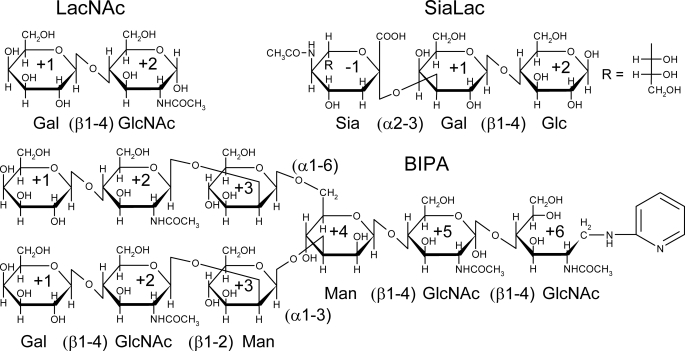
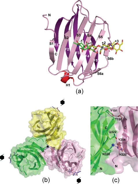
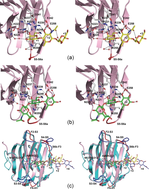
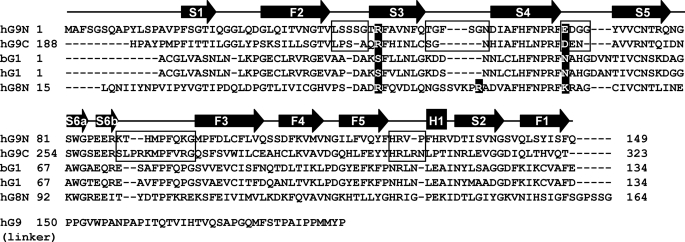
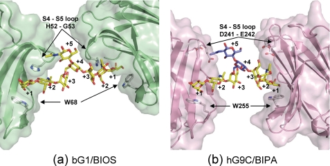
Galectin-9, a tandem-repeat-type β-galactoside-specific animal lectin with two carbohydrate recognition domains (CRDs) at the N- and C-terminal ends, is involved in chemoattraction, apoptosis, and the regulation of cell differentiation and has anti-allergic effects. Its ability to recognize carbohydrates is essential for its biological functions. Human galectin-9 (hG9) has high affinity for branched N-glycan-type oligosaccharides (dissociation constants of 0.16-0.70 μM) and linear β1-3-linked poly-N-acetyllactosamines (0.09-8.3 μM) and significant affinity for the α2-3-sialylated oligosaccharides (17-34 μM). Further, its N-terminal CRD (hG9N) and C-terminal CRD (hG9C) differ in specificity. To elucidate this unique feature of hG9, x-ray structures of hG9C in the free form and in complexes with N-acetyllactosamine, the biantennary pyridylaminated oligosaccharide, and α2-3-sialyllactose were determined. They are the first x-ray structural analysis of C-terminal CRD of the tandem-repeat-type galectin. The results clearly revealed the mechanism by which branched and α2-3-sialylated oligosaccharides are recognized and explained the difference in specificity between hG9N and hG9C. Based on structural comparisons with other galectins, we propose that the wide entrance for ligand binding and the shallow binding site of hG9C are favorable for branched oligosaccharides and that Arg(221) is responsible for recognizing sialylated oligosaccharides.
Figures
Similar articles
-
X-ray structure of a protease-resistant mutant form of human galectin-8 with two carbohydrate recognition domains.FEBS J. 2012 Oct;279(20):3937-51. doi: 10.1111/j.1742-4658.2012.08753.x. Epub 2012 Sep 11. FEBS J. 2012. PMID: 22913484
-
Evidence for subsites in the galectins involved in sugar binding at the nonreducing end of the central galactose of oligosaccharide ligands: sequence analysis, homology modeling and mutagenesis studies of hamster galectin-3.Glycobiology. 1998 Jan;8(1):45-57. doi: 10.1093/glycob/8.1.45. Glycobiology. 1998. PMID: 9451013
-
Galectin-8-N-domain recognition mechanism for sialylated and sulfated glycans.J Biol Chem. 2011 Apr 1;286(13):11346-55. doi: 10.1074/jbc.M110.195925. Epub 2011 Feb 2. J Biol Chem. 2011. PMID: 21288902 Free PMC article.
-
Full-length galectin-8 and separate carbohydrate recognition domains: the whole is greater than the sum of its parts?Biochem Soc Trans. 2020 Jun 30;48(3):1255-1268. doi: 10.1042/BST20200311. Biochem Soc Trans. 2020. PMID: 32597487 Review.
-
Oligosaccharide specificity of galectins: a search by frontal affinity chromatography.Biochim Biophys Acta. 2002 Sep 19;1572(2-3):232-54. doi: 10.1016/s0304-4165(02)00311-2. Biochim Biophys Acta. 2002. PMID: 12223272 Review.
Cited by
-
Structural Basis for Carbohydrate Recognition and Anti-inflammatory Modulation by Gastrointestinal Nematode Parasite Toxascaris leonina Galectin.J Biol Chem. 2016 Dec 2;291(49):25326-25338. doi: 10.1074/jbc.M116.743773. Epub 2016 Oct 14. J Biol Chem. 2016. PMID: 27742836 Free PMC article.
-
HSP70 Multi-Functionality in Cancer.Cells. 2020 Mar 2;9(3):587. doi: 10.3390/cells9030587. Cells. 2020. PMID: 32121660 Free PMC article. Review.
-
Single-cell RNA-seq reveals developmental plasticity with coexisting oncogenic states and immune evasion programs in ETP-ALL.Blood. 2021 May 6;137(18):2463-2480. doi: 10.1182/blood.2019004547. Blood. 2021. PMID: 33227818 Free PMC article.
-
Multifarious roles of sialic acids in immunity.Ann N Y Acad Sci. 2012 Apr;1253(1):16-36. doi: 10.1111/j.1749-6632.2012.06517.x. Ann N Y Acad Sci. 2012. PMID: 22524423 Free PMC article. Review.
-
Translational Implication of Galectin-9 in the Pathogenesis and Treatment of Viral Infection.Int J Mol Sci. 2017 Oct 8;18(10):2108. doi: 10.3390/ijms18102108. Int J Mol Sci. 2017. PMID: 28991189 Free PMC article. Review.
References
-
- Barondes S. H., Castronovo V., Cooper D. N., Cummings R. D., Drickamer K., Feizi T., Gitt M. A., Hirabayashi J., Hughes C., Kasai K., et al. (1994) Cell 76, 597–598 - PubMed
-
- Kasai K., Hirabayashi J. (1996) J. Biochem. 119, 1–8 - PubMed
-
- Rabinovich G. A., Baum L. G., Tinari N., Paganelli R., Natoli C., Liu F. T., Iacobelli S. (2002) Trends Immnol. 23, 313–320 - PubMed
Publication types
MeSH terms
Substances
Associated data
- Actions
- Actions
- Actions
- Actions
LinkOut - more resources
Full Text Sources
Other Literature Sources
Molecular Biology Databases
Research Materials
