

Conformation changes, N-terminal involvement, and cGMP signal relay in the phosphodiesterase-5 GAF domain
- PMID: 20861010
- PMCID: PMC2992248
- DOI: 10.1074/jbc.M110.141614
Conformation changes, N-terminal involvement, and cGMP signal relay in the phosphodiesterase-5 GAF domain
Abstract
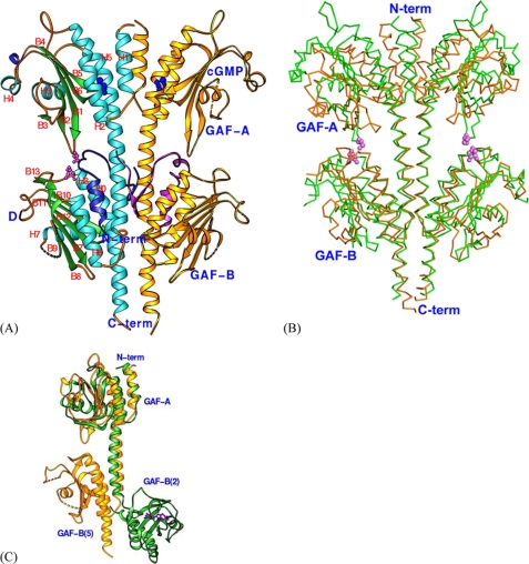
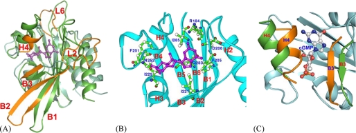
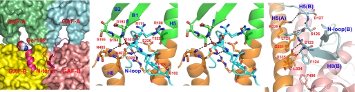
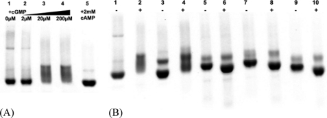
The activity of phosphodiesterase-5 (PDE5) is specific for cGMP and is regulated by cGMP binding to GAF-A in its regulatory domain. To better understand the regulatory mechanism, x-ray crystallographic and biochemical studies were performed on constructs of human PDE5A1 containing the N-terminal phosphorylation segment, GAF-A, and GAF-B. Superposition of this unliganded GAF-A with the previously reported NMR structure of cGMP-bound PDE5 revealed dramatic conformational differences and suggested that helix H4 and strand B3 probably serve as two lids to gate the cGMP-binding pocket in GAF-A. The structure also identified an interfacial region among GAF-A, GAF-B, and the N-terminal loop, which may serve as a relay of the cGMP signal from GAF-A to GAF-B. N-terminal loop 98-147 was physically associated with GAF-B domains of the dimer. Biochemical analyses showed an inhibitory effect of this loop on cGMP binding and its involvement in the cGMP-induced conformation changes.
Figures
Similar articles
-
Solution structure of the cGMP binding GAF domain from phosphodiesterase 5: insights into nucleotide specificity, dimerization, and cGMP-dependent conformational change.J Biol Chem. 2008 Aug 15;283(33):22749-59. doi: 10.1074/jbc.M801577200. Epub 2008 Jun 4. J Biol Chem. 2008. PMID: 18534985 Free PMC article.
-
The GAF domain of the cGMP-binding, cGMP-specific phosphodiesterase (PDE5) is a sensor and a sink for cGMP.Biochemistry. 2008 Mar 18;47(11):3534-43. doi: 10.1021/bi702025w. Epub 2008 Feb 23. Biochemistry. 2008. PMID: 18293931
-
Distinct allostery induced in the cyclic GMP-binding, cyclic GMP-specific phosphodiesterase (PDE5) by cyclic GMP, sildenafil, and metal ions.J Biol Chem. 2011 Mar 11;286(10):8545-8554. doi: 10.1074/jbc.M110.193185. Epub 2010 Dec 29. J Biol Chem. 2011. PMID: 21193396 Free PMC article.
-
The GAF-tandem domain of phosphodiesterase 5 as a potential drug target.Handb Exp Pharmacol. 2011;(204):151-66. doi: 10.1007/978-3-642-17969-3_6. Handb Exp Pharmacol. 2011. PMID: 21695639 Review.
-
Phosphodiesterase 5 (PDE5): Structure-function regulation and therapeutic applications of inhibitors.Biomed Pharmacother. 2021 Feb;134:111128. doi: 10.1016/j.biopha.2020.111128. Epub 2020 Dec 18. Biomed Pharmacother. 2021. PMID: 33348311 Review.
Cited by
-
Coevolving residues distant from the ligand binding site are involved in GAF domain function.Commun Chem. 2025 Apr 7;8(1):107. doi: 10.1038/s42004-025-01447-9. Commun Chem. 2025. PMID: 40195517 Free PMC article.
-
Biosynthesis of the Pseudomonas aeruginosa Extracellular Polysaccharides, Alginate, Pel, and Psl.Front Microbiol. 2011 Aug 22;2:167. doi: 10.3389/fmicb.2011.00167. eCollection 2011. Front Microbiol. 2011. PMID: 21991261 Free PMC article.
-
Advances in targeting cyclic nucleotide phosphodiesterases.Nat Rev Drug Discov. 2014 Apr;13(4):290-314. doi: 10.1038/nrd4228. Nat Rev Drug Discov. 2014. PMID: 24687066 Free PMC article. Review.
-
Structural Analysis of the Regulatory GAF Domains of cGMP Phosphodiesterase Elucidates the Allosteric Communication Pathway.J Mol Biol. 2020 Oct 2;432(21):5765-5783. doi: 10.1016/j.jmb.2020.08.026. Epub 2020 Sep 6. J Mol Biol. 2020. PMID: 32898583 Free PMC article.
-
Distinct patterns of compartmentalization and proteolytic stability of PDE6C mutants linked to achromatopsia.Mol Cell Neurosci. 2015 Jan;64:1-8. doi: 10.1016/j.mcn.2014.10.007. Epub 2014 Nov 11. Mol Cell Neurosci. 2015. PMID: 25461672 Free PMC article.
References
-
- Bender A. T., Beavo J. A. (2006) Pharmacol. Rev. 58, 488–520 - PubMed
-
- Omori K., Kotera J. (2007) Circ. Res. 100, 309–327 - PubMed
-
- Lugnier C. (2006) Pharmacol. Ther. 109, 366–398 - PubMed
-
- Conti M., Beavo J. (2007) Annu. Rev. Biochem. 76, 481–511 - PubMed
-
- Rotella D. P. (2002) Nat. Rev. Drug Discov. 1, 674–682 - PubMed
Publication types
MeSH terms
Substances
Associated data
- Actions
- Actions
Grants and funding
LinkOut - more resources
Full Text Sources
