. 2010 Dec 10;285(50):38987-9000.
doi: 10.1074/jbc.M110.175182. Epub 2010 Sep 22.

The transcriptional repressor ID2 can interact with the canonical clock components CLOCK and BMAL1 and mediate inhibitory effects on mPer1 expression

Affiliations

The transcriptional repressor ID2 can interact with the canonical clock components CLOCK and BMAL1 and mediate inhibitory effects on mPer1 expression

Sarah M Ward et al. J Biol Chem. .

Abstract

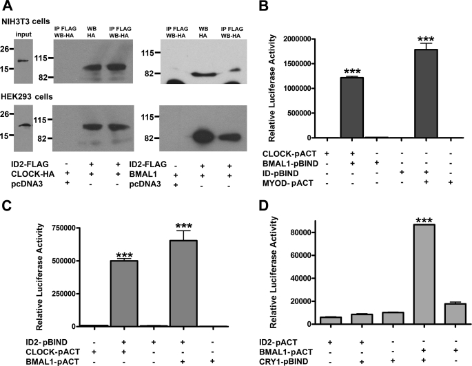
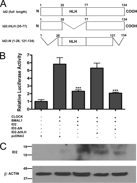
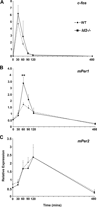
ID2 is a rhythmically expressed HLH transcriptional repressor. Deletion of Id2 in mice results in circadian phenotypes, highlighted by disrupted locomotor activity rhythms and an enhanced photoentrainment response. ID2 can suppress the transactivation potential of the positive elements of the clock, CLOCK-BMAL1, on mPer1 and clock-controlled gene (CCG) activity. Misregulation of CCGs is observed in Id2(-/-) liver, and mutant mice exhibit associated alterations in lipid homeostasis. These data suggest that ID2 contributes to both input and output components of the clock and that this may be via interaction with the bHLH clock proteins CLOCK and BMAL1. The aim of the present study was to explore this potential interaction. Coimmunoprecipitation analysis revealed the capability of ID2 to complex with both CLOCK and BMAL1, and mammalian two-hybrid analysis revealed direct interactions of ID2, ID1 and ID3 with CLOCK and BMAL1. Deletion of the ID2 HLH domain rendered ID2 ineffective at inhibiting CLOCK-BMAL1 transactivation, suggesting that interaction between the proteins is via the HLH region. Immunofluorescence analysis revealed overlapping localization of ID2 with CLOCK and BMAL1 in the cytoplasm. Overexpression of CLOCK and BMAL1 in the presence of ID2 resulted in a significant reduction in their nuclear localization, revealing that ID2 can sequester CLOCK and BMAL1 to the cytoplasm. Serum stimulation of Id2(-/-) mouse embryonic fibroblasts resulted in an enhanced induction of mPer1 expression. These data provide the basis for a molecular mechanism through which ID2 could regulate aspects of both clock input and output through a time-of-day specific interaction with CLOCK and BMAL1.


Figures

FIGURE 1.
FIGURE 1.
ID2 can interact directly with both CLOCK and BMAL1. A, NIH3T3 and HEK293 cells were cotransfected with ID2-FLAG and either CLOCK-HA or BMAL1-HA. Total cell extracts (middle lanes) or immunoprecipitates (IP, outer lanes) using an anti-FLAG antibody were subjected to Western blot (WB) analysis using anti-HA antiserum. Preimmunoprecipitation inputs for the respective immunoprecipitates are indicated (left lanes), representing 10% of input proteins and showing the presence of ID2-FLAG. ID2-FLAG signal was not detected in the negative control (not shown). Representative results are shown from one experiment. All co-immunoprecipitations were repeated at least twice in both cell lines. B–D, NIH3T3 cells were co-transfected with pGL5-Luc, β-gal, and the indicated expression plasmids. Shown are positive controls of known robustly interacting binding partners, CLOCK and BMAL1 (34, 46) and ID1 and MyoD (60) (B) and testing ID2 interaction with CLOCK and BMAL1 (C). D, as a negative control, the ability for ID2 to interact with CRY1 was tested, and a positive interaction signal was observed for BMAL1 with CRY1 (46). Luciferase reporter measurements were normalized to β-gal signals. Data are mean ± S.E. for four samples per treatment group. Differences determined by one-factor ANOVA with Bonferroni post hoc t tests. ***, p < 0.001. GAL4, DNA binding domain (pBIND plasmid); VP16, activation domain (pACT plasmid). Data are representative of three independent experiments.
FIGURE 2.
FIGURE 2.
ID1 and ID3 can interact with both CLOCK and BMAL1. A–D, NIH3T3 cells were co-transfected with pGL5-Luc, β-gal, and the indicated expression plasmids. Interaction between ID1 with CLOCK and with BMAL1 was tested using alternative ID1 plasmids with different vector backbones (A and B). Interaction between ID3 with CLOCK and with BMAL1 was also tested using two different ID3 plasmids (C and D). As a negative control, the ability for ID1 and ID3 to interact with CRY1 was tested (A and C). ID plasmids in A and C were pBIND, and ID plasmids in B and D were pGAL4. A positive interaction signal was observed for BMAL1 with CRY1 (see Fig. 1D). Luciferase reporter measurements were normalized to β-gal signals. Data are mean ± S.E. for four samples per treatment group. Differences determined by one factor ANOVA with Bonferroni post hoc t tests, *, p < 0.05; **, p < 0.01; and ***, p < 0.001. GAL4, DNA binding domain (pBIND or pGAL4 plasmid); VP16, activation domain (pACT or pVP16 plasmid). Data are representative of three independent experiments.
FIGURE 3.
FIGURE 3.
The helix-loop-helix domain of ID2 is the protein-binding site necessary for inhibition of CLOCK-BMAL1 transactivation. The effect of co-transfection of CLOCK and BMAL1 with ID2 expression plasmids on transactivation of the mPer1 promoter. NIH3T3 cells were co-transfected with mPer1:luciferase promoter reporter, β-gal, and the indicated expression plasmids. A, amino acid maps of full-length and truncated ID2 proteins encoded by DNA plasmids, with deleted residues noted in parentheses. B, mean ± S.E. luciferase reporter measurements normalized to β-gal, with five samples per treatment group. All groups, except pcDNA3.1 empty vector, were transfected with equal amount of CLOCK and BMAL1 expression vector (125 ng each). Values obtained for cells transfected with empty vector were set to 1.0. Differences determined by one-factor ANOVA with Bonferroni post hoc t tests. ***, p < 0.001 versus CLOCK-BMAL1-pcDNA3 group. Data are representative of four independent experiments. Results for inhibition of transativation by full-length ID2 is consistent with previous results (27). C, Western blot analysis of transfected cell lysates revealing protein bands for full-length ID2, ID2-ΔHLH, and ID2-ΔN truncated plasmids. Plasmid combinations for each lane are as described above in B.
FIGURE 4.
FIGURE 4.
Co-immunofluorescence reveals cytoplasmic localization of ID2 with both CLOCK and BMAL1. HEK293 cells were co-transfected with ID2 and CLOCK-HA or ID2 and BMAL1. Fluorescent dye-labeled secondary antibodies were used to visualize the cellular location of each protein. A, both the localization and co-staining signals of ID2 (green) and CLOCK (red) predominantly occur in the cytoplasm. C, both the localization and co-localization of ID2 (green) and BMAL1 (red) predominantly occurs in the cytoplasm. Cells were visualized using anti-ID2, anti-HA (for CLOCK), and anti-BMAL1 antibodies. Scale bars, 20 μm. B and D, quantification of localization data are mean ± S.E. for three independent experiments each comprising of ≥100 cell counts. The higher proportion of cells showing nuclear localization for BMAL1 compared with CLOCK is as expected from published reports (30).
FIGURE 5.
FIGURE 5.
ID2 restricts CLOCK and BMAL1 nuclear localization. HEK293 cells were co-transfected with either CLOCK-HA and BMAL1 (CB state), or CLOCK-HA, BMAL1, and ID2-FLAG (ICB state). A, CLOCK and BMAL1 localization (green and red, respectively) and co-staining (yellow) when overexpressed together and in the absence of overexpressed ID2. B, CLOCK and BMAL1 localization and co-staining in the presence of overexpressed ID2. Cellular protein localizations were visualized using anti-HA (for CLOCK) and anti-BMAL1 antibodies. Scale bar, 20 μm. C–E, quantification of localization data are mean ± S.E. for three independent experiments each comprising ≥100 cell counts. CB, ectopic expression of CLOCK and BMAL1 plasmids; ICB, ectopic expression of ID2, CLOCK, and BMAL1.
FIGURE 6.
FIGURE 6.
Serum-induced mPer1 gene expression is elevated in Id2−/− mouse embryonic fibroblasts. Serum stimulation of MEFs was used as a model system to mimic photic stimulation of the SCN. A, relative expression of c-fos in Id2−/− compared with WT (Id2+/+) MEFs shows no significant difference between genotypes. B, mPer1 expression is 2-fold higher in the peak level of gene expression in Id2−/− cells at 60 min post-initiation of serum treatment. C, relative expression of mPer2 shows no significant difference between cells. Values are mean ± S.E. fold change of expression relative to the lowest expression value from five independent experiments. c-fos, mPer1, and mPer2 expression was assessed by qRT-PCR using SYBR green and normalized to GAPDH assessed using Taqman reagents. Two-tailed Student's t test was performed at individual time points to determine statistical significance between WT and Id2−/− samples. **, p value < 0.01.
FIGURE 7.
FIGURE 7.
Proposed model for the role of ID2 in clock resetting. A, serum stimulation of MEF or light activation of retinal photoreceptors, retinohypothalamic tract (RHT), and in turn, SCN neuron (1, 5, 6, 16, 42). Intracellular signaling pathways are activated culminating in the phosphorylation of CREB on Ser-133 and Ser-142 and permitting CREB binding to its response element, CRE, in the promoter region of the immediate early gene/clock gene mPer1. This promotes transcription of the target gene (12, 13, 61). The CRE and E-box elements are located in the promoter regions of the mPer1 gene. CLOCK-BMAL1 heterodimer binding to the E-box element within the gene promoter is considered responsible for rhythmic transcriptional activity in various clock and clock-controlled genes (12, 13). Studies in the Clock Δ19 homozygous mutant mouse suggest an additional role of this binding: that CLOCK-BMAL1 binding to the E-box element is permissive to, or gates, transcriptional activation by the CRE within the same promoter region (19, 20). Target gene (mPer1 and mPer2) induction by activation of the CRE is reduced in the Clock (Clk/Clk) mutant mouse SCN and MEFs following light and serum stimulation, respectively. mPer1 is a state variable of the clock and its induction or suppression is deemed critical to phase shifting of the clock (1, 2, 10, 42). B, in the wild type cell, the presence of ID2 interferes with the dimerization of CLOCK-BMAL1 by binding to both canonical clock proteins. ID2-CLOCK and ID2-BMAL1 cannot bind to the E-box element because ID2 lacks a basic DNA binding domain. The quantity of the available CLOCK-BMAL1 heterodimer is reduced, leading to reduced transcription of the target gene mPer1. C, in the Id2−/− cell, the number of CLOCK-BMAL1 heterodimers is increased due to the absence of ID2, thereby increasing the occurrence of binding of CLOCK-BMAL1 to the E-box element and permitting maximal activation of the promoter when there is coincidental binding of pCREB to the CRE. This results in enhanced transcription of the target gene mPer1. As mPer1 is a state variable of the clock, a larger phase shift of the clock is produced (27). Additionally, a direct role for CLOCK in signaling to the mPer1 gene has also been proposed independent of the pCREB pathway, in which Ca2+ -dependent protein kinase C phosphorylation of CLOCK can regulate mPer1 induction (62). Sequestration of CLOCK to the cytoplasm and reduction in the quantities of available CLOCK-BMAL1 heterodimer by ID2 could modulate this proposed signaling pathway.

References

    1. Reppert S. M., Weaver D. R. (2001) Annu. Rev. Physiol. 63, 647–676 - PubMed
    1. Dunlap J. C., Loros J. J., DeCoursey P. J. (2004) Chronobiology: Biological Timekeeping, Sinauer Associates, Sunderland, MA
    1. Yoo S. H., Yamazaki S., Lowrey P. L., Shimomura K., Ko C. H., Buhr E. D., Siepka S. M., Hong H. K., Oh W. J., Yoo O. J., Menaker M., Takahashi J. S. (2004) Proc. Natl. Acad. Sci. U.S.A. 101, 5339–5346 - PMC - PubMed
    1. Peirson S. N., Butler J. N., Duffield G. E., Takher S., Sharma P., Foster R. G. (2006) Biochem. Biophys. Res. Commun. 351, 800–807 - PubMed
    1. Balsalobre A., Damiola F., Schibler U. (1998) Cell 93, 929–937 - PubMed

Publication types