. 2010 Nov 26;285(48):37271-80.
doi: 10.1074/jbc.M110.123471. Epub 2010 Sep 22.

A unique redox-sensing sensor II motif in TorsinA plays a critical role in nucleotide and partner binding

Affiliations

A unique redox-sensing sensor II motif in TorsinA plays a critical role in nucleotide and partner binding

Li Zhu et al. J Biol Chem. .

Abstract

Early onset dystonia is commonly associated with the deletion of one of a pair of glutamate residues (ΔE302/303) near the C terminus of torsinA, a member of the AAA+ protein family (ATPases associated with a variety of cellular activities) located in the endoplasmic reticulum lumen. The functional consequences of the disease-causing mutation, ΔE, are not currently understood. By contrast to other AAA+ proteins, torsin proteins contain two conserved cysteine residues in the C-terminal domain, one of which is located in the nucleotide sensor II motif. Depending on redox status, an ATP hydrolysis mutant of torsinA interacts with lamina-associated polypeptide 1 (LAP1) and lumenal domain like LAP1 (LULL1). Substitution of the cysteine in sensor II diminishes the redox-regulated interaction of torsinA with these substrates. Significantly, the dystonia-causing mutation, ΔE, alters the ability of torsinA to mediate the redox-regulated interactions with LAP1 and LULL1. Limited proteolysis experiments reveal redox- and mutation-dependent changes in the local conformation of torsinA as a function of nucleotide binding. These results indicate that the cysteine-containing sensor II plays a critical role in redox sensing and the nucleotide and partner binding functions of torsinA and suggest that loss of this function of torsinA contributes to the development of DYT1 dystonia.


Figures

FIGURE 1.
FIGURE 1.
Predicted structure, featured motifs, and essential functional residues of human torsinA. A, position of conserved cysteines (red), residues required for nucleotide binding and hydrolysis (green and magenta), and mutated in dystonia (blue and cyan). B, close-up of the ATPase active site of the second AAA+ domain (D2) of E. coli ClpA structure (Protein Data Bank 1ksf). The conserved ATP-binding and hydrolytic residues are shown in green and magenta, respectively, as shown in A. Two conserved cysteines that form a disulfide bond in torsinA are placed on the model using a multispecies alignment (29) and are highlighted by red spheres. The conserved Arg702 in the sensor II motif of ClpA is shown as a red stick. The positions of dystonia-associated mutations of torsinA, deletion of Glu302/303, and deletion of amino acids 323–328, are shown in different shades of blue. The ADP observed in the ClpA active site is shown in blue. The residues in parentheses are those in human torsinA.
FIGURE 2.
FIGURE 2.
The effects of the dystonia-causing mutation and the RRS mutation on the stability of torsinA. A, diagram of different versions of torsinA for stability studies, including WT, the C280S/C319S mutant (CS), the ΔE mutant (ΔE), and the ΔE with the CS mutant (ΔE/CS) of torsinA. B and C, steady state level of wild type torsinA and mutants in HeLa (B) and COS-7 (C) cells. Supernatant fractionation of cells expressing the WT, the CS mutant, the ΔE mutant, and the ΔE/CS mutant of torsinA digested with peptide:N-glycosidase F (PNGase F) or endoglycosidase H (Endo H) and immunoblotted with anti-HA antibody is shown. Molecular weight markers are shown on the left of images. Immunoblot of endogenous actin is the loading control. D, pulse-chase analysis of WT torsinA, the CS mutant, and the ΔE mutant in HeLa cells. Left panel, images from phosphorimaging; Right panel, plots of the remaining signals of the glycosylated bands. IB, immunoblot; IP, immunoprecipitation.
FIGURE 3.
FIGURE 3.
ATP-dependent interaction of torsinA with the lumenal domains of LAP1 and LULL1. A, diagram of different mutations of torsinA for the interaction studies, including WT, the E171Q mutant (EQ), and a series of mutations on the EQ background (see Fig. 4). B, ATP binding promotes the interaction between torsinA and LAP1. Cell lysates expressing WT or EQ-torsinA were treated with apyrase prior to supplementation with ATPγS, AMPPNP, ADP, ATP, or buffer and then subjected to pulldown assays. Input, 5% of cell lysate used for pulldown. IB, immunoblot.
FIGURE 4.
FIGURE 4.
The redox-sensitive cysteine in the RRS is required for the interaction between torsinA and LAP1. A, blockage of redox sensing decreases the interaction between torsinA and LAP1. Cell lysates expressing the EQ-torsinA was first treated with DTT, GSH, or buffer and then incubated with NEM for 1 mm DTT-treated sample prior to the pulldown assays. B, the interaction between torsinA and LAP1 is decreased by oxidative stress. Cells expressing the EQ-torsinA were first treated with 0.2 or 1 mm H2O2 and then lysed for the pulldown assays. C, mutation of the sensor II motif in torsinA interferes with the interaction of torsinA with LAP1. Cell lysates expressing EQ, EQ/C319S, EQ/C319A, and EQ/K320R of torsinA were subjected to the pulldown assays. D, the two cysteine residues in the C-terminal domain of torsinA, but not the N-terminal cysteines, are critical for the interaction between torsinA and LAP1. Cell lysates expressing EQ, EQ/C49S/C50S, EQ/C44S/C162S, EQ/CS, and EQ/4CS of torsinA were subjected to the pulldown assays. EQ+NEM represents the cell lysate expressing EQ-torsinA treated by NEM only prior to the pulldown assay. IB, immunoblot.
FIGURE 5.
FIGURE 5.
The dystonia-causing mutation impairs the interaction between torsinA and the lumenal domains of LAP1 and LULL1. A, diagram of different mutations of torsinA for the interaction studies, including wild type (WT), the ΔE mutant (ΔE), and a series of mutations on the ΔE or the EQ background. B, the interaction between torsinA and LAP1 is dramatically reduced by introducing the ΔE mutation into the EQ-torsinA. Cell lysates expressing WT, EQ, ΔE, ΔE/EQ, ΔE/CS, and ΔE/KA of torsinA were subjected to pulldown assays by His/Smt3–237LAP1. C, the interaction between torsinA and LAP1 as well as LULL1 is also reduced by another natural mutation on DYT1 gene, Δ323–328, but is unaffected by the missense mutation at Glu302 or Glu303. Cell lysates expressing EQ, EQ/ΔE, EQ/E302A, EQ/E303A, EQ/T324V, and EQ/Δ323–328 of torsinA were subjected to the pulldown assays by His/Smt3–237LAP1 and His/Smt3–236LULL1. IB, immunoblot.
FIGURE 6.
FIGURE 6.
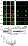
The dystonia-causing mutation alters the nucleotide-dependent conformational change in torsinA. A and B, effects of mutation on the cellular localization of torsinA. Representative immunofluorescence images of HA-tagged torsinA or mutants transiently expressed in HeLa (A) and COS-7 (B) cells and probed by anti-HA antibody. Green shows torsinA, and red shows laminB (NE marker). C, dystonia-causing mutation and mutation of the RRS of torsinA have increased sensitivity to protease. Cell lysates expressing WT, ΔE, CS, ΔE/CS, EQ, EQ/ΔE, EQ/E302A, EQ/E303A, EQ/C319A, and EQ/CS of torsinA were subjected to trypsin digestion. D, the relative amount of uncleaved protein in comparison with the control (sample without trypsin) was quantified by densitometry of the immunoblots as in C using ImageJ. The values are the means of three independent experiments with the error bars representing S.E. *, statistically significant different from WT torsinA or EQ-torsinA at p < 0.05. IB, immunoblot.
FIGURE 7.
FIGURE 7.
The dystonia-causing mutation interferes with the nucleotide binding and redox sensing activities of torsinA. A, dystonia-causing mutation and mutation of the RRS decrease the nucleotide binding activity of torsinA. Cell lysates expressing WT, ΔE, CS, EQ, EQ/ΔE, and EQ/CS of torsinA were treated with apyrase (Apy) prior to supplementation with ATPγS or buffer and then subjected to trypsin digestion. B, the relative amount of uncleaved protein in comparison with the control was quantified by densitometry of the immunoblots as in A using ImageJ. The values are the means of three independent experiments with an error bar representing S.E. *, **, ***, statistically significant different from control (sample without any treatment) at p < 0.05, p < 0.01, and p < 0.001, respectively. ATPγS stabilization for EQ-torsinA is statistically significant at p < 0.01. C, dystonia-causing mutation and mutation of the RRS lose the redox sensing activity of torsinA. Cell lysates expressing WT, ΔE, CS, EQ, EQ/ΔE, and EQ/CS of torsinA were treated with DTT or NEM or DTT and subsequent NEM, prior to trypsin digestion. D, the relative amount of uncleaved protein in comparison with the control was quantified by densitometry of the immunoblots as in C. The details are the same as in B. NS, not statistically significant. IB, immunoblot.

Similar articles

Cited by

References

    1. Ozelius L. J., Hewett J. W., Page C. E., Bressman S. B., Kramer P. L., Shalish C., de Leon D., Brin M. F., Raymond D., Corey D. P., Fahn S., Risch N. J., Buckler A. J., Gusella J. F., Breakefield X. O. (1997) Nat. Genet. 17, 40–48 - PubMed
    1. Breakefield X. O., Blood A. J., Li Y., Hallett M., Hanson P. I., Standaert D. G. (2008) Nat. Rev. Neurosci. 9, 222–234 - PubMed
    1. Neuwald A. F., Aravind L., Spouge J. L., Koonin E. V. (1999) Genome Res. 9, 27–43 - PubMed
    1. Hanson P. I., Whiteheart S. W. (2005) Nat. Rev. Mol. Cell Biol. 6, 519–529 - PubMed
    1. Doyle S. M., Wickner S. (2009) Trends Biochem. Sci 34, 40–48 - PubMed

Publication types

MeSH terms