. 2010 Dec;299(6):F1308-19.
doi: 10.1152/ajprenal.00423.2010. Epub 2010 Sep 22.

AMP-activated protein kinase inhibits KCNQ1 channels through regulation of the ubiquitin ligase Nedd4-2 in renal epithelial cells

Affiliations

AMP-activated protein kinase inhibits KCNQ1 channels through regulation of the ubiquitin ligase Nedd4-2 in renal epithelial cells

Rodrigo Alzamora et al. Am J Physiol Renal Physiol. 2010 Dec.

Abstract

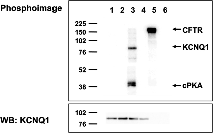
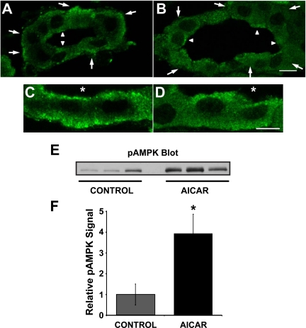
The KCNQ1 K(+) channel plays a key role in the regulation of several physiological functions, including cardiac excitability, cardiovascular tone, and body electrolyte homeostasis. The metabolic sensor AMP-activated protein kinase (AMPK) has been shown to regulate a growing number of ion transport proteins. To determine whether AMPK regulates KCNQ1, we studied the effects of AMPK activation on KCNQ1 currents in Xenopus laevis oocytes and collecting duct epithelial cells. AMPK activation decreased KCNQ1 currents and channel surface expression in X. laevis oocytes, but AMPK did not phosphorylate KCNQ1 in vitro, suggesting an indirect regulatory mechanism. As it has been recently shown that the ubiquitin-protein ligase Nedd4-2 inhibits KCNQ1 plasma membrane expression and that AMPK regulates epithelial Na(+) channels via Nedd4-2, we examined the role of Nedd4-2 in the AMPK-dependent regulation of KCNQ1. Channel inhibition by AMPK was blocked in oocytes coexpressing either a dominant-negative or constitutively active Nedd4-2 mutant, or a Nedd4-2 interaction-deficient KCNQ1 mutant, suggesting that Nedd4-2 participates in the regulation of KCNQ1 by AMPK. KCNQ1 is expressed at the basolateral membrane in mouse polarized kidney cortical collecting duct (mpkCCD(c14)) cells and in rat kidney. Treatment with the AMPK activators AICAR (2 mM) or metformin (1 mM) reduced basolateral KCNQ1 currents in apically permeabilized polarized mpkCCD(c14) cells. Moreover, AICAR treatment of rat kidney slices ex vivo induced AMPK activation and intracellular redistribution of KCNQ1 from the basolateral membrane in collecting duct principal cells. AICAR treatment also induced increased ubiquitination of KCNQ1 immunoprecipitated from kidney slice homogenates. These results indicate that AMPK inhibits KCNQ1 activity by promoting Nedd4-2-dependent channel ubiquitination and retrieval from the plasma membrane.


Figures

Fig. 1.
Fig. 1.
AMP-activated protein kinase (AMPK) inhibits KCNQ1 and KCNQ1/E1 channels expressed in Xenopus laevis oocytes. cRNAs encoding KCNQ1 alone or KCNQ1 along with KCNE1 were injected into X. laevis oocytes 3 days before the performance of two-electrode voltage clamp (TEV) experiments, as described in materials and methods. A: representative current-time sweeps with voltage-clamp steps between −100 and +100 mV are shown in oocytes expressing KCNQ1 alone (top) or KCNQ1+KCNE1 (bottom) after microinjection with either K+-gluconate (left) or K+-5-aminoimidazole-4-carboxamide-1-β-d-ribofuranosyl 5′-monophosphate (ZMP; right) 1–4 h before the measurements. B: representative chromanol 293B-sensitive current-voltage (I-V) curves are shown for oocytes expressing KCNQ1 alone (●) or KCNQ1+KCNE1 (○) that were previously microinjected with K+-gluconate control solution. C: summary of mean ± SE chromanol 293B-sensitive currents measured at +100-mV potential in oocytes expressing KCNQ1 alone (left) or KCNQ1 + KCNE1 (right) following microinjection with either K+-gluconate control (gray bars) or the AMPK activator K+-ZMP (black bars). *P < 0.01, by ANOVA for the indicated comparisons; n = 20–26 oocytes for each condition from 3 separate batches.
Fig. 2.
Fig. 2.
AMPK does not directly phosphorylate KCNQ1 in vitro. HEK-293T cells transfected to express either KCNQ1 (lanes 1–4) or CFTR (lanes 5 and 6) were lysed, and then the channels were immunoprecipitated from cell lysates for in vitro phosphorylation assays using either no added kinase (lanes 2, 4, and 6), purified AMPK holoenzyme (lanes 1 and 5), or purified PKA catalytic subunit (lane 3), as described in materials and methods. Representative phospho-screen (top) and Western blot (WB; bottom) images of the same nitrocellulose membrane are shown from 1 of 3 repeat experiments. Positive controls confirmed that AMPK was able to phoshorylate CFTR (lane 5), and PKA was able to phosphorylate KCNQ1 (lane 3), as previously described (34, 49). However, there was no detectable KCNQ1 phosphorylation by AMPK in vitro (lane 1). Western blot analysis confirmed comparable KCNQ1 expression in lanes 1–4 (bottom).
Fig. 3.
Fig. 3.
AMPK-dependent regulation of KCNQ1 channels is mediated via functional regulation of Nedd4-2. Two-electrode voltage clamp (TEV) experiments were performed on oocytes expressing KCNQ1 (wild-type or mutant) and KCNE1 along with or without xNedd4-2 (wild-type or mutant). The oocytes were then microinjected with either the AMPK activator ZMP (black bars) or K+-gluconate (KG) control (gray bars) 1–4 h before experimentation, and chromanol 293B-sensitive KCNQ1/E1 currents were measured by TEV at a voltage of +100 mV. A, left: ZMP significantly reduced KCNQ1/E1 currents compared with KG-treated oocytes. Right: expression of KCNQ1 with a PY-motif mutation (Y662A) significantly increased KCNQ1/E1 currents and prevented ZMP-dependent current inhibition. NS, not significant. B: coexpression of wild-type xNedd4-2 (1.5 ng cRNA/oocyte) inhibited KCNQ1/E1 currents, and ZMP treatment inhibited KCNQ1/E1 currents in both the presence and absence of exogenous Nedd4-2 coexpression. C: coexpression of ubiquitin-ligase activity-deficient xNedd4-2 mutant (C938S; 5 ng/oocyte) enhanced KCNQ1/E1 currents and prevented inhibition of these currents by ZMP. DN, dominant negative. D: coexpression of a constitutively active (CA) Nedd4-2 mutant (S338A/S444A; 0.7 ng) dramatically reduced KCNQ1/E1 currents and prevented inhibition of these currents by ZMP. For each condition 22–30 oocytes were recorded. *P < 0.05 by ANOVA for indicated comparison.
Fig. 4.
Fig. 4.
AMPK activation reduces plasma membrane expression of KCNQ1/KCNE1 channels. A: KCNQ1 tagged with an extracellular c-Myc epitope was coexpressed with KCNE1 subunits in X. laevis oocytes. The oocytes were then microinjected with either ZMP (black bars) or K+-gluconate control (gray bars) and incubated for 3 h. The c-myc epitope inserted in the S1-S2 extracellular loop of KCNQ1 enabled the detection of channel surface expression by a chemiluminescence assay. Activation of AMPK with ZMP significantly reduced the surface expression of KCNQ1/KCNE1 channels by ∼40–45% compared with oocytes injected with K+-gluconate. Oocytes injected with water instead of cRNA (white bar) were tested to determine background signal, which accounted for <10% of control values. To pool data from different batches of oocytes, surface expression values were normalized to the corresponding mean values of wild-type control oocytes. Values are means ± SE of 34–38 oocytes for each condition from 2 separate batches of oocytes, *P < 0.01 by unpaired t-test. B: chromanol 293B-sensitive currents were determined in parallel in oocytes from the same experimental groups. Values are means ± SE of 10–12 oocytes for each condition from 2 separate batches of oocytes, *P < 0.01 by unpaired t-test. C: representative KCNQ1 Western blot analysis showing no significant difference in whole cell expression of KCNQ1 protein between oocytes treated with ZMP or K+-gluconate.
Fig. 5.
Fig. 5.
KCNQ1 and KCNE1 expression in mouse kidney homogenates and mpkCCDc14 cells (A) and KCNQ1 cellular localization in polarized mpkCCDc14 cells (B). A, left: 30 μg of mouse total kidney homogenate (lane 1), or 50 μg of nonpolarized (lane 2) or polarized (lane 3) mpkCCDc14 cell lysates were immunoblotted with antibodies against either KCNQ1 (top), KCNE1 (middle), or β-actin (bottom), as described in materials and methods. Right: parallel samples run on the same membrane were incubated with the KCNQ1 or KCNE1 antibodies along with 10-fold excess of immunizing peptide. B: polarized mpkCCDc14 cells grown on Transwell filters were immunostained with TOPRO nuclear stain (green), KCNQ1 (red), and the tight junction marker zonula occludens (ZO)-1 (blue) as described in materials and methods. Pseudocoloring was performed for each fluorophore to enhance clarity of the images. Representative x-y sections are shown at the level of the tight junction (row A) and at a midsection of the monolayer (row B). Merged images are shown on the right. Bottom: x-z reconstruction of merged images demonstrates KCNQ1 in a basolateral membrane distribution. Scale bar = 10 μm.
Fig. 6.
Fig. 6.
AMPK activation inhibits basolateral KCNQ1 currents in polarized mpkCCDc14 cells. A: representative Western blot of cell lysates showing 24-h time course of AMPK activity (pThr172-α1-AMPK; top) with β-actin as loading control (bottom) following treatment of polarized mpkCCDc14 cells with 1 mM metformin. B: summary of means ± SE relative AMPK activities (normalized to control at time 0) following treatment for up to 24 h with 2 mM AICAR (○), 1 mM metformin (●), or vehicle control (◊). *P < 0.05, unpaired t-test compared with control at time 0; n = 3 replicate experiments for each drug treatment data point; n = 6 for controls. C: representative short-circuit current (Isc) tracings of mpkCCDc14 cells pretreated for 16 h with 1 mM metformin (dark gray trace) or vehicle (light gray trace) in the presence of an apical-to-basolateral K+ gradient, as described in materials and methods. Cell monolayers were mounted in Ussing chambers and treated first with 20 μM amiloride apically to inhibit ENaC, then 20 μM amphotericin B apically to isolate conductance at the basolateral membrane, then 50 μM of the KCNQ1 activator L-364,373 basolaterally, then 50 μM chromanol 293B basolaterally, and finally 2 mM BaCl2 basolaterally at the indicated times. One-second transepithelial voltage pulses of ±2 mV were performed each minute throughout the recordings but were removed from the displayed current trace for clarity. D: summary of means ± SE basolateral KCNQ1-mediated Isc (defined as difference between peak current following L-364,373 treatment and the current measured after chromanol 293B treatment) with or without 1 mM metformin pretreatment for 16 h. E: summary of means ± SE basolateral KCNQ1-mediated Isc with or without 2 mM AICAR pretreatment for 16 h. *P < 0.05, unpaired t-test, compared with control; n = 8 filters analyzed in 4 separate experiments for all conditions. Inset: representative immunoblots of cell lysates for pThr172-α1-AMPK and β-actin following the AICAR treatment experiment.
Fig. 7.
Fig. 7.
AICAR induces a redistribution of KCNQ1 away from the basolateral pole in collecting duct principal cells of rat kidney slices. Rat kidney slices were treated for 75 min in either control Ringer buffer alone (A and C) or Ringer buffer+2 mM AICAR (B and D) before immunofluorescence staining and laser-scanning confocal microscopy. Principal cells were identified by costaining with the principal cell marker aquaporin-2 (AQP2) or by the absence of the intercalated cell marker V-ATPase (not shown). In A and B, large arrows outside tubules indicate principal cells, and small arrowheads within tubular lumens indicate intercalated cells (scale bar = 20 μm). C and D are higher magnification images of principal cells (scale bar = 10 μm). Asterisk (*) denotes lumen. These images are representative of at least 3 separate experiments using 3 different kidney slice preparations. Three separate kidney slice lysates were immunoblotted for the active AMPK using an antibody raised against AMPK-α-pTh172 (E). Values are relative means ± SE of AMPK-α-pThr172 signal for 3 separate slices normalized to β-actin (F). *P < 0.05, unpaired t-test.
Fig. 8.
Fig. 8.
AMPK activation stimulates ubiquitination of KCNQ1 channels in rat kidney slices. Rat kidney slices were treated for 75 min in either control Ringer buffer alone of Ringer buffer containing 2 mM AICAR before lysis, homogenization, immunoprecipitation (IP), and immunoblotting, as described in materials and methods. A: representative immunoblot showing increased ubiquitination of KCNQ1 in rat kidney slices treated with AICAR compared with kidney slices treated with control buffer (top right). Ubiquitination of KCNQ1 produced a shift in the observed molecular weight of KCNQ1, consistent with the conjugation of ubiquitin chains. The amount of KCNQ1 protein in the immunoprecipitate was determined by reprobing with an anti-KCNQ1 antibody (bottom right). No ubiquitin or KCNQ1 signals were detected in parallel samples incubated without anti-KCNQ1 antibody during the immunoprecipitation. Kidney slice lysate samples were probed for KCNQ1 and β-actin (left) to assess basal expression. B: densitometric analysis of ubiquitinated KCNQ1 normalized to the corresponding amount of KCNQ1 immunoprecipitated from lysates of kidney slices treated with AICAR or control buffer. Values are means ± SE relative to control values of 4 independent kidney slice preparations. *P < 0.01 by paired t-test.

References

    1. Abriel H, Staub O. Ubiquitylation of ion channels. Physiology (Bethesda) 20: 398–407, 2005 - PubMed
    1. Acharya P, Beckel J, Ruiz WG, Wang E, Rojas R, Birder L, Apodaca G. Distribution of the tight junction proteins ZO-1, occludin, and claudin-4, -8, and -12 in bladder epithelium. Am J Physiol Renal Physiol 287: F305–F318, 2004 - PubMed
    1. Apodaca G, Katz LA, Mostov KE. Receptor-mediated transcytosis of IgA in MDCK cells is via apical recycling endosomes. J Cell Biol 125: 67–86, 1994 - PMC - PubMed
    1. Bacallao R, Stelzer EH. Preservation of biological specimens for observation in a confocal fluorescence microscope and operational principles of confocal fluorescence microscopy. Methods Cell Biol 31: 437–452, 1989 - PubMed
    1. Belfodil R, Barriere H, Rubera I, Tauc M, Poujeol C, Bidet M, Poujeol P. CFTR-dependent and -independent swelling-activated K+ currents in primary cultures of mouse nephron. Am J Physiol Renal Physiol 284: F812–F828, 2003 - PubMed

Publication types