. 2010 Dec;84(23):12200-9.
doi: 10.1128/JVI.01633-10. Epub 2010 Sep 22.

A double mutation in glycoprotein gB compensates for ineffective gD-dependent initiation of herpes simplex virus type 1 infection

Affiliations

A double mutation in glycoprotein gB compensates for ineffective gD-dependent initiation of herpes simplex virus type 1 infection

Hiroaki Uchida et al. J Virol. 2010 Dec.

Abstract

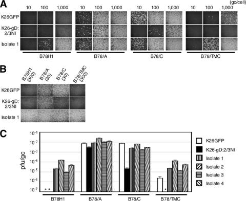
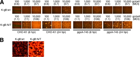
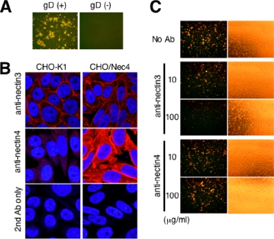
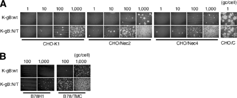
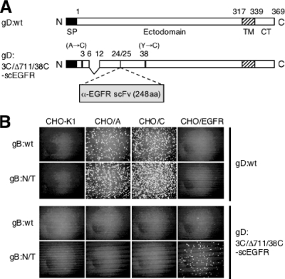
Herpes simplex virus (HSV) entry into cells is triggered by the binding of envelope glycoprotein D (gD) to a specific receptor, such as nectin-1 or herpesvirus entry mediator (HVEM), resulting in activation of the fusion effectors gB and gH and virus penetration. Here we report the identification of a hyperactive gB allele, D285N/A549T, selected by repeat passage of a gD mutant virus defective for nectin-1 binding through cells that express a gD-binding-impaired mutant nectin-1. The gB allele in a wild-type virus background enabled the use of other nectins as virus entry receptors. In addition, combination of the mutant allele with an epidermal growth factor receptor (EGFR)-retargeted gD gene yielded dramatically increased EGFR-specific virus entry compared to retargeted virus carrying wild-type gB. Entry of the gB mutant virus into nectin-1-bearing cells was markedly accelerated compared to that of wild-type virus, suggesting that the gB mutations affect a rate-limiting step in entry. Our observations indicate that ineffective gD activation can be complemented by hypersensitization of a downstream component of the entry cascade to gD signaling.


Figures

FIG. 1.
FIG. 1.
Tropisms of the selected virus isolates. (A) Cells were infected with isolate 1 or controls for 8 h at the various gc/cell indicated above the panels and immunostained for VP16. Infections at 1,000 gc/cell were performed separately. (B) Cells were infected with isolate 1 for 6 h at 30 or 300 gc/cell, as indicated in parentheses, and immunostained for ICP4. (C) Biological titers (PFU/ml) were divided by genome titers (gc/ml), and the mean values ± standard deviations (SD) from three determinations were plotted on a logarithmic scale. *, <10−7.
FIG. 2.
FIG. 2.
Effect of cellular glycosaminoglycans on K-gB:N/T infectivity. (A) Cells were infected for 8 or 24 h at the various gc/cell indicated above the panels and immunostained for VP16. MOIs (PFU/cell) based on virus titers on B78/C cells (PFU/ml) are included in parentheses. (B) Higher-magnification images of 10,000-gc/cell infections of CHO-K1 cells at 8 hpi.
FIG. 3.
FIG. 3.
Roles of gD and nectin-3 in infection of CHO-K1 cells by K-gB:N/T. (A) CHO-K1 cells were infected with K-gB:N/T (left panel) or K-gB:N/TΔgD (right panel) for 16 h at 1,000 gc/cell and immunostained for VP16. (B) CHO-K1 or CHO/Nec4 cells were immunostained for nectin-3 or nectin-4 and observed under a confocal microscope. Cells that reacted with the secondary antibody (Ab) alone are shown at the bottom. Nuclei were stained with Hoechst 33342. (C) CHO-K1 cells were preincubated with PBS or isotype-matched anti-nectin-3 or anti-nectin-4 monoclonal antibodies at 10 or 100 μg/ml; incubated with K-gB:N/T at 3,000 gc/cell for 2 h, followed by acidic buffer treatment; and immunostained for VP16 at 16 hpi. The panels to the right show phase-contrast images of the same fields.
FIG. 4.
FIG. 4.
Effects of gB:N/T on infection through weak gD receptors. (A) Cells were infected for 24 h at the various gc/cell indicated above the panels and immunostained for VP16. (B) Cells were infected for 8 h at 100 or 1,000 gc/cell and immunostained for VP16.
FIG. 5.
FIG. 5.
Effects of gB:N/T on EGFR-specific infection by transiently retargeted HSV. (A) Schematic representation of gD:wt and retargeted gD:3C/Δ711/38C-scEGFR. SP, signal peptide; TM, transmembrane region; CT, cytoplasmic domain; α-EGFR scFV, anti-EGFR single-chain antibody; aa, amino acids. (B) Vero cells were transfected with expression plasmids for the gD proteins indicated to the right and then infected with K-gB:wtΔgD or K-gB:N/TΔgD (indicated to the left) at MOIs of 5, followed by treatment with acidic buffer. Equal volumes of supernatant collected the next day were used for infection of the cells indicated at the top. Cells were immunostained for ICP4 at 6 hpi.
FIG. 6.
FIG. 6.
Effects of gB:N/T on virus entry kinetics. B78/C cells (A) or Vero cells (B) were incubated with 200 PFU of K-gB:wt or K-gB:N/T at 4°C for 30 min, washed thoroughly, incubated at 37 or 30°C for the indicated times, and treated with acidic buffer. Cells were then incubated at 37°C under methylcellulose-containing medium for 2 to 3 days to allow plaque formation. Plaques were counted, and the mean values ± SD from three determinations were plotted. temp., temperature. (C) Cells were incubated with K-gB:wt or K-gB:N/T at 4°C for 30 min at 0.3 PFU/cell (B78/C) or for 1 h at 1 PFU/cell (CHO/C), washed thoroughly, incubated at 37°C for the various times indicated above the panels, and treated with acidic buffer. Cells were then incubated at 37°C for 8 h and immunostained for VP16. (D) Vero cells were transfected with expression plasmids for the gB genes indicated to the left, infected with KΔT, and treated with acidic buffer. Fresh Vero monolayers were then incubated with equal volumes of the respective supernatants at 4°C for 30 min, and the kinetics of virus entry were examined as in panel C. (E) Entry kinetics on CHO/A cells. Virus input was 3 PFU/cell, and the experiment was performed as in panel C, with minor modifications.

Similar articles

Cited by

References

    1. Anderson, D. B., S. Laquerre, K. Ghosh, H. P. Ghosh, W. F. Goins, J. B. Cohen, and J. C. Glorioso. 2000. Pseudotyping of glycoprotein D-deficient herpes simplex virus type 1 with vesicular stomatitis virus glycoprotein G enables mutant virus attachment and entry. J. Virol. 74:2481-2487. - PMC - PubMed
    1. Arii, J., M. Uema, T. Morimoto, H. Sagara, H. Akashi, E. Ono, H. Arase, and Y. Kawaguchi. 2009. Entry of herpes simplex virus 1 and other alphaherpesviruses via the paired immunoglobulin-like type 2 receptor alpha. J. Virol. 83:4520-4527. - PMC - PubMed
    1. Asano, R., Y. Sone, K. Makabe, K. Tsumoto, H. Hayashi, Y. Katayose, M. Unno, T. Kudo, and I. Kumagai. 2006. Humanization of the bispecific epidermal growth factor receptor x CD3 diabody and its efficacy as a potential clinical reagent. Clin. Cancer Res. 12:4036-4042. - PubMed
    1. Atanasiu, D., J. C. Whitbeck, T. M. Cairns, B. Reilly, G. H. Cohen, and R. J. Eisenberg. 2007. Bimolecular complementation reveals that glycoproteins gB and gH/gL of herpes simplex virus interact with each other during cell fusion. Proc. Natl. Acad. Sci. U. S. A. 104:18718-18723. - PMC - PubMed
    1. Avitabile, E., C. Forghieri, and G. Campadelli-Fiume. 2007. Complexes between herpes simplex virus glycoproteins gD, gB, and gH detected in cells by complementation of split enhanced green fluorescent protein. J. Virol. 81:11532-11537. - PMC - PubMed

Publication types

MeSH terms