. 2010 Dec;84(23):12405-18.
doi: 10.1128/JVI.01415-10. Epub 2010 Sep 22.

Cellular immediate-early gene expression occurs kinetically upstream of Epstein-Barr virus bzlf1 and brlf1 following cross-linking of the B cell antigen receptor in the Akata Burkitt lymphoma cell line

Affiliations

Cellular immediate-early gene expression occurs kinetically upstream of Epstein-Barr virus bzlf1 and brlf1 following cross-linking of the B cell antigen receptor in the Akata Burkitt lymphoma cell line

Jianjiang Ye et al. J Virol. 2010 Dec.

Abstract

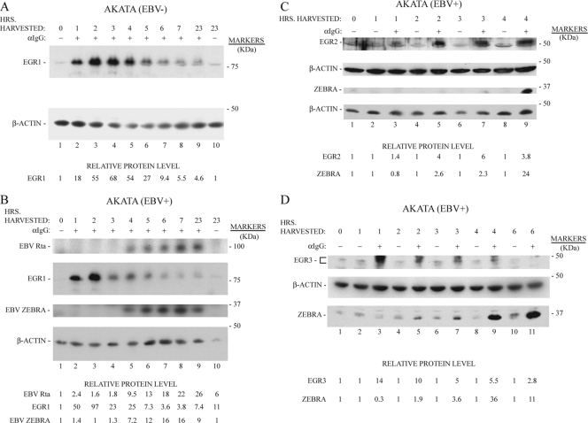
The Epstein-Barr virus (EBV) lytic activator genes bzlf1 and brlf1 are conventionally referred to as immediate-early (IE) genes. However, previous studies showed that the earliest expression of these genes was blocked by cycloheximide when the EBV lytic cycle was induced by histone deacetylase (HDAC) inhibitors and protein kinase C agonists. Anti-IgG activates a complex signal transduction pathway that leads to EBV lytic activation in the Akata cell line. Here we demonstrate that in Akata cells, where lytic cycle activation occurs very rapidly after anti-IgG treatment, de novo protein synthesis is also required for induction of bzlf1 and brlf1 expression. New protein synthesis is required up to 1.25 h after application of anti-IgG; bzlf1 and brlf1 mRNAs can be detected 1.5 h after anti-IgG. Five cellular IE genes were shown to be expressed by 1 h after addition of anti-IgG, and their expression preceded that of bzlf1 and brlf1. These include early growth response genes (egr1, egr2, and egr3) and nuclear orphan receptors (nr4a1 and nr4a3). These genes were activated by anti-IgG treatment of Akata cells with and without the EBV genome; therefore, their expression was not dependent on expression of any EBV gene product. EGR1, EGR2, and EGR3 proteins were kinetically upstream of ZEBRA and Rta proteins. Expression of EGR1, ZEBRA, and Rta proteins were inhibited by bisindolylmaleimide X, a selective inhibitor of PKC. The findings suggest a revised model in which the signal transduction cascade activated by cross-linking of the B cell receptor induces expression of cellular IE genes, such as early growth response and nuclear orphan receptor genes, whose products, in turn, regulate bzlf1 and brlf1 expression.


Figures

FIG. 1.
FIG. 1.
Kinetics of expression of EBV brlf1 and bzlf1 mRNA in Akata cells treated with anti-IgG. Akata cells were treated with anti-IgG or untreated. One aliquot of anti-IgG-exposed and control cells was also treated continuously with cycloheximide (CHX). Samples collected at the indicated times were analyzed for brlf1 (A) and bzlf1 (B) mRNA by qRT-PCR. The stimulation index (S.I.) is the ratio of brlf1 or bzlf1 mRNA in the anti-IgG-treated samples relative to that of control samples collected at the same time.
FIG. 2.
FIG. 2.
Inhibitors of protein synthesis block activation of EBV brlf1 and bzlf1 expression in Akata cells treated with anti-IgG. Akata cells were untreated (−) or treated (+) with anti-IgG. Cycloheximide (CHX) or anisomycin (ANIS) were added for the indicated intervals of time (HRS., hours). brlf1 (Rp) and bzlf1 (Zp) mRNAs were analyzed by Northern blotting. Total RNA from each sample was harvested at 8 h (panel A) or 3 h (panel B) after addition of anti-IgG.
FIG. 3.
FIG. 3.
Kinetics of inhibition of anti-IgG induction of bzlf1 and brlf1 expression by cycloheximide. Akata cells were untreated (−) or treated (+) with anti-IgG. CHX was added to individual cultures at time zero or at 15-min intervals thereafter. Total RNA was harvested from each culture at 2 h and analyzed for brlf1 (A) and bzlf1 (B) mRNA by qRT-PCR. Stimulation index (S.I.) is the ratio of brlf1 or bzlf1 mRNA in the anti-IgG-treated samples relative to that in samples that were not treated with anti-IgG. When CHX was present, the uninduced control cells were exposed to CHX for the same duration and RNA was harvested at the same time as the anti-Ig plus CHX sample. % S.I. refers to the stimulation index of sample pairs that were exposed to CHX for the indicated time normalized to that of the sample pair that was not exposed to CHX, set at 100%.
FIG. 4.
FIG. 4.
Effect of cycloheximide on the level of six cellular mRNAs whose expression was induced by treatment of Akata cells with anti-IgG. The experiment was conducted with EBV-positive and -negative Akata cells. The cells were untreated (−) or treated (+) with anti-IgG. One culture in each group was treated with CHX. The levels of egr1 (A), egr2 (B), egr3 (C), nab2 (D), nr4A1 (E), and nr4a3 (F) cellular mRNAs were compared with those of brlf1 (G) and bzlf1 (H) using qRT-PCR. All the samples were harvested after 2.5 h.
FIG. 5.
FIG. 5.
Kinetics of cellular gene mRNA expression relative to bzlf1 and brlf1 in Akata cells treated with anti-IgG. Akata cells were untreated (−) or treated (+) with anti-IgG (αIgG). Total RNA harvested at the times indicated was analyzed for cellular and viral gene expression by qRT-PCR.
FIG. 6.
FIG. 6.
Kinetics of EGR1, EGR2, and EGR3 protein expression in Akata cells treated with anti-IgG. EBV-negative (A) and EBV-positive (B, C, and D) Akata cells were untreated (−) or treated (+) with anti-IgG (αIgG). Cell extracts prepared at the times indicated were analyzed for expression of EGR1 protein (A and B), EGR2 protein (C), or EGR3 protein (D) and of EBV Rta, ZEBRA, and β-actin by immunoblotting. The relative protein level was determined by densitometry and corrected for the β-actin level.
FIG. 7.
FIG. 7.
Kinetics of NAB2 protein expression in Akata cells treated with anti-IgG. EBV-negative (A) or EBV-positive (B and C) Akata cells were untreated (−) or treated (+) with anti-IgG (αIgG). Cell extracts prepared at the times indicated were analyzed for NAB2 and EBV EA-D (BMRF1) and ZEBRA (BZLF1). The relative protein level was determined by densitometry and corrected for the β-actin level.
FIG. 8.
FIG. 8.
Induction of EGR1, NAB2, EBV Rta, and EBV ZEBRA protein expression in Akata cells treated with anti-IgG is blocked by an inhibitor of protein kinase C. Akata cells were untreated (−) or treated (+) with anti-IgG. Aliquots of each group of cells were untreated (−) or treated (+) with bisindolylmaleimide X (Bis X), a PKC inhibitor. Cell extracts prepared at time zero (0) or at 1, 2, 3, or 4 h were analyzed by immunoblotting for expression of EGR1, NAB2, EBV Rta, EBV ZEBRA, or β-actin by immunoblotting with specific antibodies. Cell extracts from the same experiment were utilized for panels A to C. In panel A, the immunoblot was developed with 125I protein 4. Panels B and C represent separate gels that were developed with ECL.
FIG. 9.
FIG. 9.
Effect of transfection of Akata cells with a mixture of plasmids encoding cellular immediate-early genes on expression of EBV bzlf1. Akata cells were cotransfected with a vector expressing CAAX-GFP and a control vector plasmid (Ctrl.) or with CAAX-GFP and a mixture of plasmids expressing 4 cellular immediate-early genes, egr1, egr2, nr4a1, and nr4a3. After 20 to 22 h cells were separated into GFP-positive and -negative populations and analyzed for bzlf1 expression by qRT-PCR. In panel A, the cells received no additional treatment. In panel B, the cells were treated with CHX 2.5 h before harvest. In panel C, the cells were treated with 7.5 μg/ml of anti-IgG for 1.5 h before harvest. In panel D, the cells were treated with 0.2 μg/ml of anti-IgG 2.5 h before harvest. The relative level of BZLF1 expression in all samples was compared to the expression level in GFP-negative cells from the population transfected with the control vector.
FIG. 10.
FIG. 10.
EGR1 induces EBV bzlf1 and brlf1 expression in 293 cells containing an EBV bacmid. 2089 cells were transfected with expression vectors for five cellular genes whose expression was induced by treatment of Akata cells with anti-IgG. mRNA harvested 48 h after transfection was analyzed for EBV bzlf1 (A) or brlf1 (B) expression by qRT-PCR.

References

    1. Adamson, A. L., D. Darr, E. Holley-Guthrie, R. A. Johnson, A. Mauser, J. Swenson, and S. Kenney. 2000. Epstein-Barr virus immediate-early proteins BZLF1 and BRLF1 activate the ATF2 transcription factor by increasing the levels of phosphorylated p38 and c-Jun N-terminal kinases. J. Virol. 74:1224-1233. - PMC - PubMed
    1. Adamson, A. L., and S. Kenney. 2001. Epstein-Barr virus immediate-early protein BZLF1 is SUMO-1 modified and disrupts promyelocytic leukemia bodies. J. Virol. 75:2388-2399. - PMC - PubMed
    1. Ben-Sasson, S. A., and G. Klein. 1981. Activation of the Epstein-Barr virus genome by 5-aza-cytidine in latently infected human lymphoid lines. Int. J. Cancer 28:131-135. - PubMed
    1. Bhaduri-McIntosh, S., and G. Miller. 2006. Cells lytically infected with Epstein-Barr virus are detected and separable by immunoglobulins from EBV-seropositive individuals. J. Virol. Methods 137:103-114. - PubMed
    1. Bhende, P. M., W. T. Seaman, H. J. Delecluse, and S. C. Kenney. 2004. The EBV lytic switch protein, Z, preferentially binds to and activates the methylated viral genome. Nat. Genet. 36:1099-1104. - PubMed

Publication types

MeSH terms

LinkOut - more resources