. 2010 Dec;84(23):12292-9.
doi: 10.1128/JVI.01700-10. Epub 2010 Sep 22.

Cascade of events governing cell-cell fusion induced by herpes simplex virus glycoproteins gD, gH/gL, and gB

Affiliations

Cascade of events governing cell-cell fusion induced by herpes simplex virus glycoproteins gD, gH/gL, and gB

Doina Atanasiu et al. J Virol. 2010 Dec.

Abstract

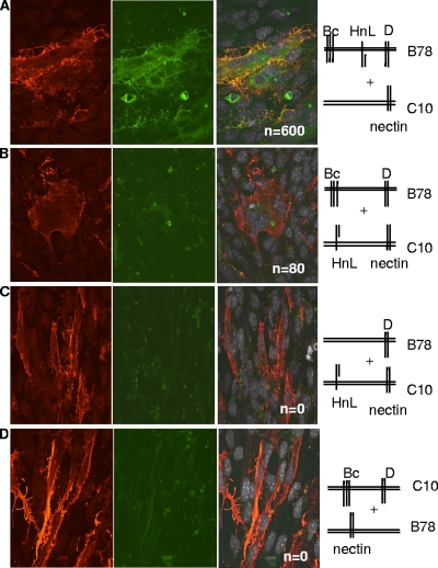
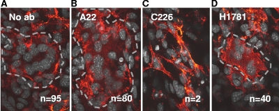
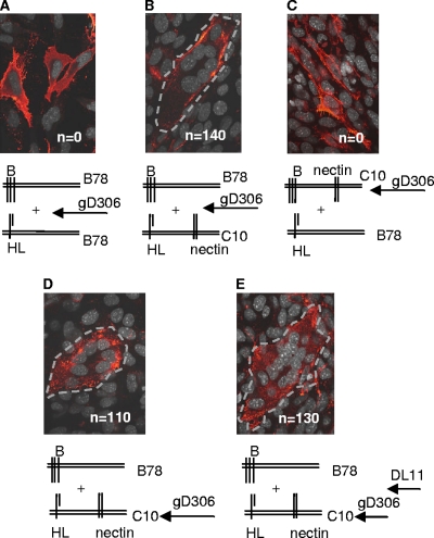
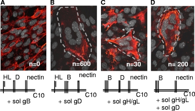
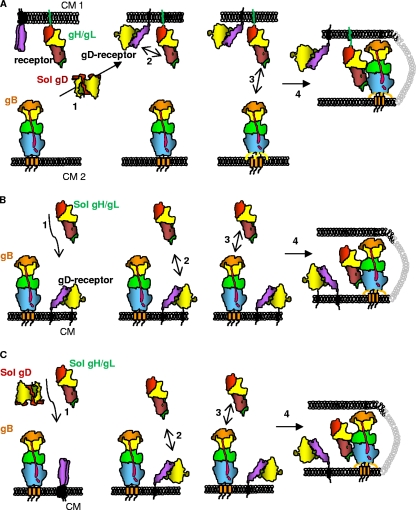
Herpesviruses minimally require the envelope proteins gB and gH/gL for virus entry and cell-cell fusion; herpes simplex virus (HSV) additionally requires the receptor-binding protein gD. Although gB is a class III fusion protein, gH/gL does not resemble any documented viral fusion protein at a structural level. Based on those data, we proposed that gH/gL does not function as a cofusogen with gB but instead regulates the fusogenic activity of gB. Here, we present data to support that hypothesis. First, receptor-positive B78H1-C10 cells expressing gH/gL fused with receptor-negative B78H1 cells expressing gB and gD (fusion in trans). Second, fusion occurred when gH/gL-expressing C10 cells preexposed to soluble gD were subsequently cocultured with gB-expressing B78 cells. In contrast, prior exposure of gB-expressing C10 cells to soluble gD did not promote subsequent fusion with gH/gL-expressing B78 cells. These data suggest that fusion involves activation of gH/gL by receptor-bound gD. Most importantly, soluble gH/gL triggered a low level of fusion of C10 cells expressing gD and gB; a much higher level was achieved when gB-expressing C10 cells were exposed to a combination of soluble gH/gL and gD. These data clearly show that gB acts as the HSV fusogen following activation by gD and gH/gL. We suggest the following steps leading to fusion: (i) conformational changes to gD upon receptor binding, (ii) alteration of gH/gL by receptor-activated gD, and (iii) upregulation of the fusogenic potential of gB following its interaction with activated gH/gL. The third step may be common to other herpesviruses.


Figures

FIG. 1.
FIG. 1.
Fusion occurs when gB and gH/gL are in trans. (A to D) Nectin-1-expressing B78H1-C10 cells and/or B78 cells were transfected for 8 h with plasmids for the glycoproteins as shown in the stick diagrams. The C10 cells were trypsinized and overlaid onto coverslips containing B78 cells, and the bilayer was cocultured for 40 h. Cells were fixed and stained with anti-gH/gL polyclonal antibody (red). Propidium iodide was added to visualize nuclei (gray). Cells were analyzed by immunofluorescent assay for protein (red channel), EYFP (green channel), and nuclei (far-red channel). Images in the far-red channel were artificially colored white, as seen in the merged images (third set of images). Confocal images are at ×60 magnification and were captured using the same camera setting. n, number of syncytia per coverslip.
FIG. 2.
FIG. 2.
Cell-cell fusion in trans is blocked by the anti-gB neutralizing MAb C226. B78H1 cells transfected with gB and gD were cocultured with C10 cells transfected with gH/gL in the absence (A) or presence (B) of the nonneutralizing MAb A22 or in the presence of the neutralizing MAb C226 (C) or H1781 (D). Conditions for coculture, fixation, and staining are the same as for Fig. 1. For easier identification, the syncytia were outlined with a dotted gray line. n, number of syncytia per coverslip.
FIG. 3.
FIG. 3.
Fusion in trans is triggered by preexposure of C10 cells expressing gH/gL to soluble gD306. (A) B78 cells expressing gB were cocultured with B78 cells expressing gH/gL. Soluble gD306 was added at the time of coculture (negative control). (B) B78 cells expressing gB were cocultured with C10 cells expressing gH/gL. Soluble gD306 was added at the time of coculture (positive control). (C) C10 cells expressing gB were incubated with gD306 (soluble gD) overnight. The medium was removed, and the cells were washed once with medium, overlaid with fresh medium and serum, and cocultured with B78 cells expressing gH/gL. (D) C10 cells expressing gH/gL were incubated with gD306 (soluble gD) overnight. The medium was removed, and the cells were washed once with medium, overlaid with fresh medium and serum, and cocultured with B78 cells expressing gB. (E) Same as in panel D, except that MAb DL11 was added at the time of coculture between the C10 and B78 cells. Dotted gray lines in panels B, D, and E outline the identified syncytia. n, number of syncytia per coverslip.
FIG. 4.
FIG. 4.
Neither gD nor gH/gL needs to be membrane anchored to trigger cell-cell fusion. (A to D) C10 cells were transfected with plasmids for the proteins indicated in the diagrams for 8 h, followed by addition of soluble gB (A), gD (B), or gH/gL (C) or a combination of gD and gH/gL (D). Panels A and B were stained for gH/gL; panels C and D were stained for gB. Syncytia are outlined for easier identification. n, number of syncytia per coverslip.
FIG. 5.
FIG. 5.
Schemes for the steps in fusion when gB is triggered by receptor-bound soluble gD and/or gH/gL. (A to C) Each protein is depicted in cartoon form, shaped like the actual structure. Glycoproteins gB and gH/gL are colored according to the color scheme used to depict their domains (12, 18). gD is colored according to the color scheme used to depict its dimeric structure (24). (A) In step 1, soluble gD binds to the cell-bound receptor (purple) and undergoes conformational changes that enable it to interact with cell-bound gH/gL (step 2). Then, cell-bound gH/gL interacts with cell-bound gB (step 3) to cause fusion (step 4). (B) Soluble gH/gL is added to cells containing gD bound to the receptor (steps 1 and 2). Then, steps 3 and 4 happen as in panel A. (C) A combination of soluble gD and gH/gL is added to receptor-bearing cells expressing gB. gD binds the receptor (step 1), undergoes conformational changes, and then acts upon gH/gL (step 2), which is then able to upregulate the fusogenic activity of gB (steps 3 and 4). CM, cell membrane.

Similar articles

Cited by

References

    1. Atanasiu, D., J. C. Whitbeck, T. M. Cairns, B. Reilly, G. H. Cohen, and R. J. Eisenberg. 2007. Bimolecular complementation reveals that glycoproteins gB and gH/gL of herpes simplex virus interact with each other during cell fusion. Proc. Natl. Acad. Sci. U. S. A. 104:18718-18723. - PMC - PubMed
    1. Atanasiu, D., J. C. Whitbeck, M. P. de Leon, H. Lou, B. P. Hannah, G. H. Cohen, and R. J. Eisenberg. 2010. Bimolecular complementation defines functional regions of herpes simplex virus gB that are involved with gH/gL as a necessary step leading to cell fusion. J. Virol. 84:3825-3834. - PMC - PubMed
    1. Avitabile, E., C. Forghieri, and G. Campadelli-Fiume. 2007. Complexes between herpes simplex virus glycoproteins gD, gB, and gH detected in cells by complementation of split enhanced green fluorescent protein. J. Virol. 81:11532-11537. - PMC - PubMed
    1. Avitabile, E., C. Forghieri, and G. Campadelli-Fiume. 2009. Cross talk among the glycoproteins involved in herpes simplex virus entry and fusion: the interaction between gB and gH/gL does not necessarily require gD. J. Virol. 83:10752-10760. - PMC - PubMed
    1. Backovic, M., and T. S. Jardetzky. 2009. Class III viral membrane fusion proteins. Curr. Opin. Struct. Biol. 19:189-196. - PMC - PubMed

Publication types

Substances

LinkOut - more resources