. 2010 Nov 15;21(22):3890-901.
doi: 10.1091/mbc.E10-06-0520. Epub 2010 Sep 22.

Paradoxical instability-activity relationship defines a novel regulatory pathway for retinoblastoma proteins

Affiliations

Paradoxical instability-activity relationship defines a novel regulatory pathway for retinoblastoma proteins

Pankaj Acharya et al. Mol Biol Cell. .

Abstract

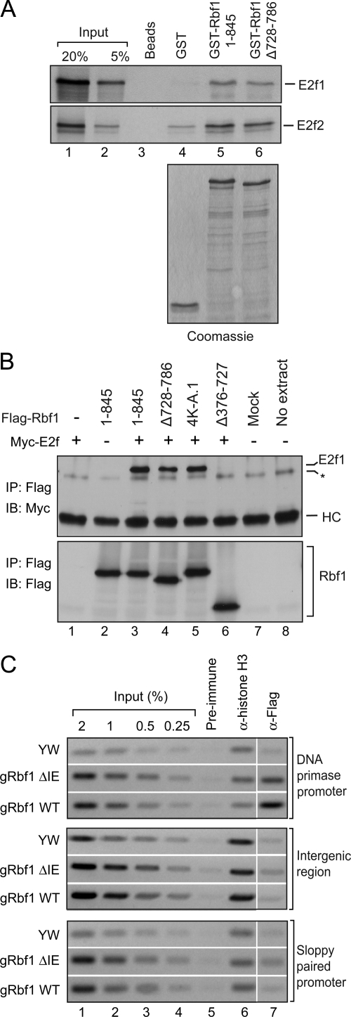
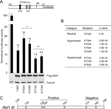
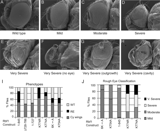
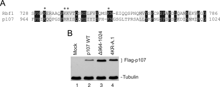
The retinoblastoma (RB) transcriptional corepressor and related family of pocket proteins play central roles in cell cycle control and development, and the regulatory networks governed by these factors are frequently inactivated during tumorigenesis. During normal growth, these proteins are subject to tight control through at least two mechanisms. First, during cell cycle progression, repressor potential is down-regulated by Cdk-dependent phosphorylation, resulting in repressor dissociation from E2F family transcription factors. Second, RB proteins are subject to proteasome-mediated destruction during development. To better understand the mechanism for RB family protein instability, we characterized Rbf1 turnover in Drosophila and the protein motifs required for its destabilization. We show that specific point mutations in a conserved C-terminal instability element strongly stabilize Rbf1, but strikingly, these mutations also cripple repression activity. Rbf1 is destabilized specifically in actively proliferating tissues of the larva, indicating that controlled degradation of Rbf1 is linked to developmental signals. The positive linkage between Rbf1 activity and its destruction indicates that repressor function is governed in a manner similar to that described by the degron theory of transcriptional activation. Analogous mutations in the mammalian RB family member p107 similarly induce abnormal accumulation, indicating substantial conservation of this regulatory pathway.


Figures

Figure 1.
Figure 1.
Identification of an instability element (IE) in Rbf1. (A) Schematic diagram of Rbf1 proteins expressed in Drosophila S2 cells. The N and C termini are indicated in dark and light gray, respectively; the black box represents the instability element; the E2f-binding pocket domain is in white. (B) Effect of proteasome inhibitor MG132 on Rbf1 protein levels. Cells were transfected to express the indicated proteins and treated for 1–8 h with MG132, and protein levels assayed by Western blot using antibodies to C-terminal Flag epitope tag. The wild-type 1-845 and mutants lacking the extreme C terminus (Δ787-845) or the pocket domain deletion mutant (Δ376-727) were expressed at lower levels and were strongly stabilized by this drug, while the mutants lacking the IE (Δ728-786 and 1-727) were expressed at higher levels and were not much further stabilized by MG132 treatment. (C) Effects of cell density and culture time on differential expression of wild-type Rbf1 and IE mutant. 400 ng of Rbf1 expression plasmid was transfected into S2 cells. At lower initial cell densities (0.75 × 10⁶/ml) and shorter growth times (3 d), expression of wild-type Rbf1 (1-845) and a deletion mutant lacking the IE (Δ 728-786) accumulate to similar levels. Normalized protein levels are shown below the lanes containing Rbf1. Cells at higher initial densities (1.5–3 × 10⁶/ml) grown for longer times (5 d) show higher levels of the mutant protein relative to the wild-type form. Levels of transfected CtBP protein, and endogenous tubulin protein, are shown as controls.
Figure 2.
Figure 2.
Conserved lysine residues in IE play critical roles in accumulation and stability of Rbf1. (A) Mutation of multiple lysine residues within the IE leads to increased protein accumulation. Lysine residues were changed to alanine (K732A, K739A, K740A for 3K-A; also K754A for 4K-A; also K774A and K782A for 6K-A) or to arginine. Rbf1 overaccumulation is not observed with the lysine to arginine substitution. 1.5 × 10⁶ S2 cells were transfected with 100 ng of Rbf1 expression plasmid and grown for five days. The data shown are representative of three biological experiments. (B and C) Half-life measurements of unstable wild-type and stable IE mutant Rbf1 proteins. Three days after transfection, cells were treated with cycloheximide and harvested at the indicated times. Rbf1 protein levels were quantitated by photon-capture analysis with a Fuji LAS-3000 Imager and normalized to tubulin levels. (D) Bar graphs showing averaged normalized flag:tubulin ratios for the Rbf1 wild-type and 4K-A mutant proteins at the 6-h time point from three biological replicates. At this time point, the difference between the wild-type and the 4K-A mutant protein levels was statistically significant (p = 0.05).
Figure 3.
Figure 3.
Expression of wild-type and IE mutant forms of Rbf1 in the Drosophila larva. Indicated proteins were expressed from the endogenous rbf1 promoter, and expression levels were assayed in total larval extracts as well as in imaginal discs. (A) Western blot showing expression of Flag-tagged Rbf1 from third-instar larvae (left panel) and pooled imaginal discs (right panel) carrying homozygous copies of rbf1 genomic constructs. Equivalent levels of proteins were noted in whole larval extracts whereas the mutant protein was found to accumulate to ∼fourfold of the wild-type protein in the imaginal discs. The Western blot of whole larval extracts is representative of four biological replicates for the two lines shown in C, F, I, and D, G, J; the average difference in protein levels in total larval extracts was 13% ± 2%. (B–J) Rbf1 expression in third-instar larval imaginal discs. (B–D) Eye discs, (E–G) wing discs, and (H–J) leg discs. Weak background staining was observed in nontransgenic yw flies (B, E, and H), and specific but weak staining was evident in discs expressing wild-type Rbf1 protein (C, F, and I). Strong expression was noted in flies expressing the inactive Rbf1 Δ 728-786 IE mutant protein (D, G, and J). The imaginal disc staining is representative of stainings of three different lines for each construct; in all cases, the IE mutant protein was expressed at higher levels.
Figure 4.
Figure 4.
Rbf1 requires the IE for transcriptional repression. (A) Deletion of the IE (Δ 728-786) or E2f binding pocket (Δ376-727) compromises transcriptional repression activity of Rbf1 proteins measured on the PCNA-luciferase reporter gene (bar graph). Under these transfection conditions, proteins were expressed at similar levels (Western blot). (B) Subcellular localization of wild-type (1–845) and deletion mutants. DAPI staining indicates DNA in nucleus, and FITC staining the Rbf1 proteins. Proteins lacking residues 787–845, which include the presumptive nuclear localization signal, are found predominantly in the cytoplasm. (C) Transcriptional activity of Rbf1 IE deletion and point mutant proteins assayed on PCNA-luciferase reporter. Mutant proteins lacking the IE, or with multiple lysine to alanine mutations, were compromised for transcriptional repression activity. Lysine to arginine mutant proteins exhibited wild-type repression activity. Error bars indicate SD, and asterisks indicate p < 0.05 (D) Rbf1 repression of Drosophila Polα-luciferase reporter. Deletion of the IE largely inactivates the protein for transcriptional repression (top panel). Data in 4A represent two biological replicates, each with three technical replicates, except for 1-845 and Δ 728-786, which represent 16 and 9 biological replicates. Other transfections include data from at least three biological replicates. Firefly luciferase activity is expressed relative to Renilla luciferase control.
Figure 5.
Figure 5.
Rbf1 IE is not essential for E2F interactions and promoter binding. (A and B) Physical association between Rbf1 IE mutants and E2F proteins. (A) GST-Rbf1 and E2f interaction assay. Indicated GST fusion proteins were bound to radio-labeled E2f proteins and bound proteins were analyzed by SDS-PAGE and autoradiography. GST-Rbf1 1-845 and ΔIE mutant displayed similar binding ability to both in vitro translated E2f1 and E2f2 proteins (compare lanes 5 and 6). No interaction was observed with beads alone and GST protein (lanes 3 and 4). Coomassie stained gel showing equal amounts of GST fusion proteins used in binding assays (bottom panel). The data shown are representative of three biological replicates. (B) Coimmunoprecipitation assay. Rbf1/E2f1 interactions in cotransfected S2 cells. Cells were cotransfected with Myc-tagged E2f1 and Flag-tagged Rbf1 expression constructs. Whole cell lysates were used for Flag immunoprecipitations (IP) and the samples were assayed using Western blots with anti-Myc antibody (top panel). Myc-tagged E2f1 coprecipitated with Rbf1 1-845 and two IE mutants (Δ728-786 and 4K-A.1) but not with the pocket domain deletion mutant (Δ376-727) (top panel, lanes 3–6). Mock is IP performed using cell lysate from untransfected cells (lane 7). The asterisk indicates a nonspecific band that is contributed by the Flag M2 beads since it appeared in the no extract control where IP was performed in the absence of any cell lysate (lane 8). Equivalent levels of the heavy chain IgG (marked as HC) were seen in all samples indicating the use of equal amount of antibody for each IP reaction. The IP samples were also blotted with the anti-Flag antibody (bottom panel) to verify the amount of Flag-tagged protein that was captured in each assay. The data shown are representative of two biological replicates. (C) Promoter occupancy by Flag-tagged Rbf1 wild-type and Rbf1 IE mutant proteins measured by chromatin immunoprecipitation. Formaldehyde cross-linked chromatin was prepared from 0 to 20 h embryos expressing the wild-type or mutant Rbf1 protein and immunoprecipitated using the indicated antibodies. Enrichment of the Rbf-regulated promoter (DNA primase) was observed by anti-Flag antibody immunoprecipitation reactions with both wild-type and IE mutant fly embryos but not in reactions using pre-immune IgG (top panel) or at an intergenic locus (middle panel) and a nontarget gene promoter (sloppy paired 1) (bottom panel).
Figure 6.
Figure 6.
Rbf1 IE harbors positive and negative regulatory elements. (A) Transcriptional repression activity of Rbf1 lysine point mutant proteins. Examples of mutant proteins that show either enhanced or reduced repression activity. Mutation of K754 to alanine or arginine attenuates repression activity while K774 to alanine mutant exhibited enhanced repression activity with respect to the wild-type protein (top panel). Under these transfection conditions, proteins were expressed at similar levels (lower panel). Error bars indicate standard deviations, and asterisks indicate p < 0.05 compared with wild-type Rbf1. (B) The lysine point mutants were classified as neutral, hypo-, or hypermorphic based on the indicated t test results. (C) Schematic representation of the Rbf1 IE indicating the location of lysine residues that play a positive or negative role in Rbf1-mediated repression.
Figure 7.
Figure 7.
Severe developmental consequences of expression of hyperactive Rbf1. cDNAs of rbf1 wild-type and IE hypermorphic and hypomorphic mutants were misexpressed in the eye imaginal disc using the eye-Gal4 driver. (A–H) representative eyes exhibiting wild-type, mild, moderate, severe, and four very severe phenotypes. (I) Bar graphs representing frequency with which flies carrying the eye-Gal4 driver and UAS-rbf1 gene were recovered, as well as frequency with which these latter flies exhibited a phenotype (“WT” normal eye, “RE” rough eye of any degree of severity, “Cy wings” indicates flies that lacked the Gal4 driver, did not express the rbf1 transgene, and had wild-type eyes). Note that Δ728-786 and 1-727, which lack the IE and were inactive in cell culture, never showed a phenotype, and that the hyperactive K774 mutants exhibited a partially lethal phenotype, as judged by lower recovery of flies containing the eye-Gal4 driver. (J) Severity of eye phenotype in flies exhibiting rough eyes. Mutants are shown in order of increasing severity; point mutations in the IE that decreased function in cell culture assays also exhibited weaker eye phenotypes, and hypermorphic K774 alleles exhibited much stronger phenotypes.
Figure 8.
Figure 8.
Mutations in the conserved IE of p107 enhance expression. (A) Similarities between Rbf1 IE and homologous region of p107, which is most similar to Rbf1. Asterisks mark basic residues mutated in each protein to stabilize expression. (B) Genes for Flag-tagged wild-type p107 or IE mutants were transfected into S2 cells and expression quantitated by Western blot. The 60-aa region deleted from p107 in Δ964-1024 is similar to the Rbf1 IE. Endogenous tubulin levels are shown as controls.

References

    1. Binne U. K., Classon M. K., Dick F. A., Wei W., Rape M., Kaelin W. G., Jr, Naar A. M., Dyson N. J. Retinoblastoma protein and anaphase-promoting complex physically interact and functionally cooperate during cell-cycle exit. Nat. Cell. Biol. 2007;9:225–232. - PubMed
    1. Boyer S. N., Wazer D. E., Band V. E7 protein of human papilloma virus-16 induces degradation of retinoblastoma protein through the ubiquitin-proteasome pathway. Cancer Res. 1996;56:4620–4624. - PubMed
    1. Brand A. H., Perrimon N. Targeted gene expression as a means of altering cell fates and generating dominant phenotypes. Development. 1993;118:401–415. - PubMed
    1. Chang E. C., Schwechheimer C. ZOMES III: the interface between signalling and proteolysis. Meeting on The COP9 Signalosome, Proteasome and eIF3. EMBO Rep. 2004;5:1041–1045. - PMC - PubMed
    1. Classon M., Harlow E. The retinoblastoma tumour suppressor in development and cancer. Nat. Rev. Cancer. 2002;2:910–917. - PubMed

Publication types

MeSH terms

Substances

LinkOut - more resources