. 2010 Nov 15;21(22):3985-97.
doi: 10.1091/mbc.E10-05-0456. Epub 2010 Sep 22.

MAL/VIP17, a new player in the regulation of NKCC2 in the kidney

Affiliations

MAL/VIP17, a new player in the regulation of NKCC2 in the kidney

Monica Carmosino et al. Mol Biol Cell. .

Abstract

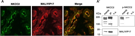
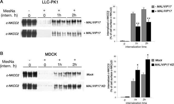
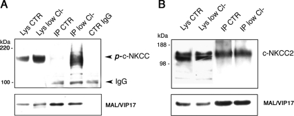
The renal-specific Na+-K+-2Cl- cotransporter (NKCC2) is the major salt transport pathway of the apical membrane of the mammalian thick ascending limb of Henle's loop. Here, we analyze the role of the tetraspan protein myelin and lymphocytes-associated protein (MAL)/VIP17 in the regulation of NKCC2. We demonstrated that 1) NKCC2 and MAL/VIP17 colocalize and coimmunoprecipitate in Lilly Laboratories cell porcine kidney cells (LLC-PK1) as well as in rat kidney medullae, 2) a 150-amino acid stretch of NKCC2 C-terminal tail is involved in the interaction with MAL/VIP17, 3) MAL/VIP17 increases the cell surface retention of NKCC2 by attenuating its internalization, and 4) this coincides with an increase in cotransporter phosphorylation. Interestingly, overexpression of MAL/VIP17 in the kidney of transgenic mice results in cysts formation in distal nephron structures consistent with the hypothesis that MAL/VIP17 plays an important role in apical sorting or in maintaining the stability of the apical membrane. The NKCC2 expressed in these mice was highly glycosylated and phosphorylated, suggesting that MAL/VIP17 also is involved in the stabilization of NKCC2 at the apical membrane in vivo. Thus, the involvement of MAL/VIP17 in the activation and surface expression of NKCC2 could play an important role in the regulated absorption of Na+ and Cl- in the kidney.


Figures

Figure 1.
Figure 1.
Colocalization and coimmunoprecipitation experiments in rat kidney medulla. (A) Sequential sections of rat kidney were stained for NKCC2 (green) and MAL/VIP17 (red). The image merge showed that NKCC2 codistributes with MAL/VIP17. (A′) Rat kidney medullae membranes were solubilized in CHAPS and subjected to immunoprecipitation using MAL/VIP17 antibody. Western blotting on kidney membranes (memb) and Immunoprecipitates (IP) showed that NKCC2 coimmunoprecipitated with MAL/VIP17 (NKCC2). A nonimmune serum (a polyclonal anti-AQP4 antibody) was used as negative control (n.s.). The same membrane was stripped and subjected to Western blotting with R5 antibody, showing that also the phosphorylated form of NKCC2 coimmunoprecipitated with MAL/VIP17 (p-NKCC2).
Figure 2.
Figure 2.
c-NKCC2 expression in LLC-PK1 and MDCK cells. (A) LLC-PK1 cells stably expressing c-NKCC2 were transfected with MAL/VIP17. Colocalization experiments were then performed using a monoclonal anti-HA antibody to stain c-NKCC2 (green) and a polyclonal anti-FLAG antibody to stain MAL/VIP17 (red). The merge showed that c-NKCC2 and MAL/VIP17 colocalize at the apical membrane. (A′) The same cells were lysed in CHAPS and subjected to immunoprecipitation with the polyclonal anti-FLAG antibody. Western blotting analysis using T4 antibody showed that c-NKCC2 coimmunoprecipitated with MAL/VIP17. The structure of the c-NKCC2 construct is shown on the right of the corresponding Western blotting. The NKCC1 regions are shown in blue, and the NKCC2 regions are shown in red. (B) Endogenously expressed MAL/VIP17 was knocked down by siRNA in c-NKCC2-transfected MDCK cells. Immunofluorescence showed that c-NKCC2 was expressed at the apical membrane in both control (MOCK) and MAL/VIP17 knocked down cells (MAL/VIP17 KD). (B′) Western blotting of MAL/VIP17 and flotillin-2 (Flo-2) in DRM fractions prepared from MDCK (MDCK DRMs). The densitometric analysis of the MAL/VIP17 bands showed a decrease of ∼70% of MAL/VIP17 expression in MAL/VIP7 knocked down cells (MAL/VIP17 densitometry). Student's t test, *p < 0.0001. (C) LLC-PK1 cells stably expressing NKCC1 were transfected with MAL/VIP17. The merge showed that NKCC1 and MAL/VIP17 did not colocalize. (C′) The same cells were lysed in CHAPS and subjected to immunoprecipitation with the polyclonal anti-FLAG antibody. Western blotting analysis using T4 antibody showed that NKCC1 did not coimmunoprecipitate with MAL/VIP17.
Figure 3.
Figure 3.
Internalization experiments. (A) LLC-PK1 cells expressing c-NKCC2 alone or in combination with MAL/VIP17 were grown to confluence and biotinylated from their apical side by using a cleavable biotin. Cells were then allowed to internalize surface-labeled proteins for the indicated times. The remaining surface accessible biotin was then stripped with MesNa where indicated (MesNa, +). Biotinylated proteins were then recovered and immunoblotted for NKCC2. Each experimental condition was examined in triplicate. Internalized c-NKCC2 was determined from three independent experiments by densitometry of biotinylated c-NKCC2 at 1 and 2 h of internalization and expressed as percentage of total cell surface c-NKCC2 (MesNa −, intern. h 0). Student's t test, **p < 0.0001. (B) Control (mock) and MAL/VIP17 knocked down (MAL/VIP17 KD) MDCK cells were subjected to the same assay described above. Each experimental condition was examined in triplicate. Internalized c-NKCC2 was determined from three independent experiments by densitometry of biotinylated c-NKCC2 at 1 and 2 h of internalization and expressed as percentage of total cell surface c-NKCC2 (MesNa −, intern. h 0). Student's t test, *p < 0.005.
Figure 4.
Figure 4.
Phosphorylation dynamics of NKCC2 in the absence or in the presence of MAL/VIP17. (A) LLC-PK1cells expressing c-NKCC2 alone or in combination with MAL/VIP17 were grown to confluence on 12-well plates, incubated in low Cl medium or in the presence of 0.5 μM CalA. Cells were then lysed and subjected to Western blotting using R5 antibody. The same nitrocellulose membranes were stripped and reprobed by Western blotting with T4 antibody. Each experimental condition was examined in quadruplicate. (B) The quantitation of phosphorylated c-NKCC2 was determined from three independent experiments by densitometry of phospho-c-NKCC2 bands (R5 signal) normalized for the corresponding c-NKCC2 bands (T4 signal). Student's t test, *p < 0.001, **p < 0.0001. (C) Both control (MOCK) and MAL/VIP17-depleted MDCK cells (MAL/VIP17 KD) were incubated in regular or low Cl medium. Cells were then lysed and subjected to Western blotting using R5 antibody (p-c-NKCC2). The same nitrocellulose membranes were stripped and reprobed by Western blotting with T4 antibody (c-NKCC2). Each experimental condition was examined in triplicate. (D) The quantitation of phosphorylated c-NKCC2 was determined from three independent experiments by densitometry of phospho-c-NKCC2 bands (p-c-NKCC2) normalized for the corresponding c-NKCC2 bands (c-NKCC2). Student's t test, **p < 0.0001.
Figure 5.
Figure 5.
Coimmunoprecipitation of c-NKCC2 and MAL/VIP17 in CTR and low Cl conditions. LLC-PK1 cells expressing c-NKCC2 and MAL/VIP17 were incubated in CTR or low Cl conditions, lysed in 2% CHAPS, and subjected to immunoprecipitatation with anti-FLAG antibody. Western blotting analysis was made with R5 (A) or T4 (B) antibodies. The phosphorylation of the cotransporter in low Cl did not change the apparent affinity of c-NKCC2 for MAL/VIP17.
Figure 6.
Figure 6.
Floatation and detergent solubility assays in LLC-PK1 cells transfected with c-NKCC2 alone or in combination with MAL/VIP17. (A) LLC-PK1 cells were lysed in 2% CHAPS at 4°C. Lysates were then subjected to floatation assay as described in Materials and Methods. Twenty-two fractions, taken from the top to the bottom of the gradient, were subjected to immunoblotting with antibodies specific for the membrane proteins indicated on the right. These data are representative of three independent experiments. The expression of MAL/VIP17 did not induce any further recruitment of c-NKCC2 into DRMs. (B) LLC-PK1 cells were lysed in 2% CHAPS at 4°C. Lysates were then fractionated into supernatant (soluble fraction) and pellet (insoluble fraction), and equal amounts of total proteins was analyzed by SDS-PAGE followed by immunoblotting with antibodies specific for the membrane proteins indicated on the right. The expression of MAL/VIP17 did not induce any further recruitment of p-c-NKCC2 into DRMs.
Figure 7.
Figure 7.
Analysis of NKCC2 and p-NKCC2 in control and MAL/VIP17-overexpressing transgenic mice. Kidneys from three control (CTR) and three transgenic (OVER-MAL/VIP17) mice were lysed in antiphosphatase buffer with 1% Triton X-100 and subjected to immunoblotting. (A) Western blotting was made with anti-NKCC2 (T4), anti-actin, and MAL/VIP17 antibodies. (A′) Densitometric analysis of the 130- to 160-kDa broad band revealed by T4 antibody, corresponding to the glycosylated form of NKCC2 (gly-NKCC2) in both strains of mice. Values were normalized for β-actin signal. (A″) Western blotting analysis was made with anti-NKCC (T4) in kidney lysates from MAL/VIP17-overexpressing transgenic mice after incubation without (−) or with (+) PNGase F. The Western blotting reported is representative of three independent experiments. (B) Western blotting was made with anti-phospho-NKCC (R5) or anti-actin antibodies. (B′) Densitometric analysis of the 160-kDa band revealed by R5 antibody, normalized for β-actin signal, in both strains of mice. The densitometric values are expressed as mean ± SE of three independent experiments. Student's t test, *p < 0.001. (C) Sections from paraformaldehyde-fixed kidneys of control or transgenic mice were subjected to immunofluorescence with R5 antibody. Images were collected with a confocal laser-scanning microscope from kidneys of three control and three transgenic mice. Two representative pictures are presented. The staining with R5 was present in cells lining the cysts (C) as well as in normal tubules (T) in MAL/VIP17-transgenic mice (OVER-MAL/VIP17). The staining is absent in kidneys from control mice.

References

    1. Alvarez-Guerra M., Garay R. P. Renal Na-K-Cl cotransporter NKCC2 in Dahl salt-sensitive rats. J. Hypertension. 2002;20:721–727. - PubMed
    1. Caplan M. J., Kamsteeg E. J., Duffield A. Tetraspan proteins: regulators of renal structure and function. Curr. Opin. Nephrol. Hypertens. 2007;16:353–358. - PubMed
    1. Carmosino M., Gimenez I., Caplan M., Forbush B. Exon loss accounts for differential sorting of Na-K-Cl cotransporters in polarized epithelial cells. Mol. Biol. Cell. 2008;19:4341–4351. - PMC - PubMed
    1. Charrin S., le Naour F., Silvie O., Milhiet P. E., Boucheix C., Rubinstein E. Lateral organization of membrane proteins: tetraspanins spin their web. Biochem. J. 2009;420:133–154. - PubMed
    1. Cheong K. H., Zacchetti D., Schneeberger E. E., Simons K. MAL/VIP17/MAL, a lipid raft-associated protein, is involved in apical transport in MDCK cells. Proc. Natl. Acad. Sci. USA. 1999;96:6241–6248. - PMC - PubMed

Publication types

MeSH terms

LinkOut - more resources