. 2010 Sep 22;30(38):12664-75.
doi: 10.1523/JNEUROSCI.1586-10.2010.

A hierarchical phosphorylation cascade that regulates the timing of PERIOD nuclear entry reveals novel roles for proline-directed kinases and GSK-3beta/SGG in circadian clocks

Affiliations

A hierarchical phosphorylation cascade that regulates the timing of PERIOD nuclear entry reveals novel roles for proline-directed kinases and GSK-3beta/SGG in circadian clocks

Hyuk Wan Ko et al. J Neurosci. .

Abstract

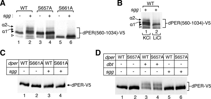
The daily timing of when PERIOD (PER) proteins translocate from the cytoplasm to the nucleus is a critical step in clock mechanisms underpinning circadian rhythms in animals. Numerous lines of evidence indicate that phosphorylation plays a prominent role in regulating various aspects of PER function and metabolism, including changes in its daily stability and subcellular distribution. In this report, we show that phosphorylation of serine 661 (Ser661) by a proline-directed kinase(s) is a key phospho-signal on the Drosophila PER protein (dPER) that regulates the timing of its nuclear accumulation. Mutations that block phosphorylation at Ser661 do not affect dPER stability but delay its nuclear entry in key pacemaker neurons, yielding longer behavioral rhythms. Intriguingly, abolishing phosphorylation at Ser661 also attenuates the extent of dPER hyperphosphorylation in vivo, suggesting the phosphorylated state of Ser661 regulates phosphorylation at other sites on dPER. Indeed, we identify Ser657 as a site that is phosphorylated by the glycogen synthase kinase GSK-3β (SHAGGY; SGG) in a manner dependent on priming at Ser661. Although not as dramatic as mutating Ser661, mutations that abolish phosphorylation at Ser657 also lead to longer behavioral periods, suggesting that a multi-kinase hierarchical phosphorylation module regulates the timing of dPER nuclear entry. Together with evidence in mammalian systems, our findings implicate proline-directed kinases in clock mechanisms and suggest that PER proteins are key downstream targets of lithium therapy, a potent inhibitor of GSK-3β used to treat manic depression, a disorder associated with clock malfunction in humans.


Figures

Figure 1.
Figure 1.
Phosphorylation of S661 on dPER, which is highly conserved in Drosophila, regulates the extent of DBT-mediated hyperphosphorylation but not the stability of dPER. A, Sequence alignment of dPER proteins from different Drosophila species (** identifies the conserved Ser–Pro pair; S661–P662 in D. melanogaster). Sequences were aligned using the program ClustalX (www.clustal.org) and processed with GeneDoc (www.nrbsc.org/gfx/genedoc) for better visualization. Amino acids that are identical in all the species analyzed are highlighted in black, whereas those showing less conservation are indicated in gray. Note that the conserved Ser–Pro pair falls in a relatively nonconserved region of dPER. B, C, S2 cells were transiently transfected with pAct–dper(560–1034)–V5–His plasmid that contained either the wild-type (WT) control version (B, lane 1; C) or the S661A mutant (B, lane 2). The wild-type dPER(560–1034) protein exhibits at least two mobility isoforms (indicated at left, arrowheads). C, Immunoprecipitated dPER(560–1034) was mock treated (lane 1) or treated with λ-phosphatase (λPPase) in the absence (lane 2) or presence (lane 3) of the phosphatase inhibitor Na3VO4. D, E, S2 cells were transiently cotransfected with pMT–dbt–V5–His and plasmids containing full-length versions of either wild-type dper (WT) or the S661A mutant (SA). Cells were collected at the indicated times after dbt induction (top) and dPER–V5 analyzed either directly by immunoblotting (D, lanes 1–6; E, lanes 1–4) or after immunoprecipitation and treatment with λ-phosphatase (D, lanes 7, 8). Where indicated, the proteasome inhibitor MG132 (50 μm final) and the translation inhibitor cycloheximide (CHX; 10 μg/ml final) were added to the media at 20 h after dbt induction, and cells were collected 4 h later. Each experiment was done at least three times, and representative examples are shown.
Figure 2.
Figure 2.
GSK-3β/SGG phosphorylates S657 and likely other sites on dPER in a hierarchical manner that depends on previous phosphorylation of S661. A, S2 cells were transiently cotransfected with the indicated versions of pAct--dper(560–1034)–V5 [wild type (WT); S657A or S661A] and pMT–sgg–V5–His. Cells were collected either just before inducing recombinant sgg (−) or 24 h after sgg induction (+). Recombinant dPER was visualized by immunoblotting in the presence of anti-V5 antibodies. Exogenous expression of sgg leads to the appearance on several novel mobility isoforms of wild-type dPER(560–1034), herein termed α1 and α2 (arrows, left). Also indicated are the S661 phosphorylated (filled arrowhead) and S661 nonphosphorylated (open arrowhead) dPER(560–1034) mobility isoforms observed in the absence of exogenously expressed kinases (see Fig. 1B). Note that the S657A mutant does not block S661 phosphorylation (lane 4), and, in the presence of recombinant SGG, only the α2 isoform is detected (lane 4). In the case of the S661A mutant, both the α1 and α2 isoforms are not detected (lane 6). B, Detection of the SGG-mediated α1 and α2 dPER(560–1034) isoforms is blocked in the presence of the GSK-3β/SGG inhibitor LiCl (10 mm, final; lane 2) but not KCl (10 mm, final; lane 1). Recombinant sgg was induced for 24 h. C, D, S2 cells were transiently cotransfected with plasmids containing different versions of full-length dper–V5–His [wild type (WT); S657A or S661A] and either pMT–sgg–V5–His or pMT–dbt–V5–His, as indicated (−, +). Cells were collected 24 h after induction of dbt or sgg, and dPER–V5 was visualized by immunoblotting. Each experiment was done at least three times, and representative examples are shown.
Figure 3.
Figure 3.
Phosphorylation of S661 by MAPK can prime additional phosphorylation at S657 by GSK-3β. A, B, Wild-type (WT), S661A, or S657A versions of dPER(560–1034)–V5–His were immunoprecipitated from extracts prepared from S2 cells using anti-V5 agarose and pretreated with λ-phosphatase for 30 min, followed by extensive washing and equilibration in MAPK kinase assay buffer. Subsequently, the immune complexes were incubated with (+) or without (−) 500 U of ERK2. After this incubation, immune complexes were washed with extraction buffer, equilibrated in GSK-3β buffer, and incubated with (+) or without (−) 500 U of GSK-3β. Phosphorylation was detected by adding [γ-32P]ATP to the in vitro kinase assays (see Materials and Methods), and radiolabeled bands were visualized by PAGE and autoradiography. The different phospho-isoforms are indicated (arrows, right of panels) and whether Ser657 or Ser661 are phosphorylated (bold). Note that ERK2 does not require previous phosphorylation to phosphorylate S661 of dPER (A, lane 3), an event that can prime subsequent phosphorylation by GSK-3β (A, lane 4). Addition of GSK-3β by itself does not lead to noticeable mobility shift in dPER(560–1034) (A, compare lanes 1 and 2). Addition of ERK2 does not shift the mobility of the S661A mutant (B, compare lanes 1 and 3), and the addition of GSK-3β after ERK2 does not evoke an additional shift in the electrophoretic mobility of either the S661A mutant (B, compare lanes 3 and 4) or S657A mutant (B, compare lanes 5 and 6). Each experiment was done at least three times, and representative examples are shown.
Figure 4.
Figure 4.
S661 is phosphorylated in flies and modulates the hyperphosphorylation of dPER but does not have significant effects on its overall daily levels. A–D, Adult flies of the indicated genotype in the wper0 genetic background (top of panels) were collected at the indicated time during the fourth day of LD (A, C, D) or first day of DD (B). A–C, Head extracts were prepared and analyzed by immunoblotting using anti-HA antibodies. CT, Circadian time. D, Head extracts were subjected to immunoprecipitation using anti-HA antibodies. A portion of the recovered immune complexes was analyzed by immunoblotting in the presence of either anti-HA (top panel) or S661 phospho-specific (anti-pS661; bottom panel) antibodies. For lanes 7 and 8, immune complexes were first treated with phosphatase before analysis by immunoblotting; note that, whereas phosphatase treatment abolishes the ability of the anti-pS661 antibodies to detect dPER (bottom panel, compare lanes 7 and 8 with 1 and 5, respectively), it still reacts with the anti-HA antibody (top panel, lanes 7 and 8). Each experiment was done at least three times, and representative examples are shown.
Figure 5.
Figure 5.
Cycling of dper mRNA levels in p{dper/S661A} flies. A, B, Transgenic flies expressing either a wild-type (WT; wper0;p{dper}) or the S661A mutant (wper0;p{dper/S661A}) version of dper were collected at the indicated times during either the fourth day of LD (A) or first day of DD (B). The relative levels of dper RNA were measured. Results from at least two independent experiments were pooled, and the error bars indicate SEM.
Figure 6.
Figure 6.
The timing of when dPER(S661A) enters the nucleus of key pacemaker neurons is delayed. Transgenic flies expressing either a wild-type (WT; wper0;p{dper}) or S661 mutant (S661A; wper0;p{dper/S661A}) version of dper were collected at the indicated times in LD (A) or at ZT22 (B, C) and processed for whole-mount immunohistochemistry. Shown are representative staining patterns obtained for the small LNvs. A, Two independent examples are shown for the S661A mutant. B, C, For each genotype, the subcellular localization of dPER at ZT22 was determined for 24 small LNvs from at least five flies; three representative examples are shown for each genotype (B), and the staining distributions for all 24 small LNvs were tabulated (C). dPER was visualized with anti-HA antibodies (shown in green). PDF was visualized with an anti-PDF antibody (shown in red) and serves as a convenient cytoplasmic marker for the small LNvs. Note that, whereas punctate nuclear staining of control dPER is clearly observed by ZT22, the subcellular localization of dPER(S661A) is much more dispersed at this time with primarily cytoplasmic or mixed cytoplasmic–nuclear staining, eventually exhibiting primarily nuclear staining by ZT24.

Similar articles

Cited by

References

    1. Akten B, Jauch E, Genova GK, Kim EY, Edery I, Raabe T, Jackson FR. A role for CK2 in the Drosophila circadian oscillator. Nat Neurosci. 2003;6:251–257. - PubMed
    1. Bae K, Edery I. Regulating a circadian clock's period, phase and amplitude by phosphorylation: insights from Drosophila. J Biochem. 2006;140:609–617. - PubMed
    1. Bao S, Rihel J, Bjes E, Fan JY, Price JL. The Drosophila double-timeS mutation delays the nuclear accumulation of period protein and affects the feedback regulation of period mRNA. J Neurosci. 2001;21:7117–7126. - PMC - PubMed
    1. Chang DC, Reppert SM. A novel C-terminal domain of Drosophila PERIOD inhibits dCLOCK:CYCLE-mediated transcription. Curr Biol. 2003;13:758–762. - PubMed
    1. Chiu JC, Vanselow JT, Kramer A, Edery I. The phospho-occupancy of an atypical SLIMB-binding site on PERIOD that is phosphorylated by DOUBLETIME controls the pace of the clock. Genes Dev. 2008;22:1758–1772. - PMC - PubMed

Publication types

MeSH terms

Substances

LinkOut - more resources