. 2010 Sep 22;30(38):12676-89.
doi: 10.1523/JNEUROSCI.1243-10.2010.

Fibrillar amyloid-beta-activated human astroglia kill primary human neurons via neutral sphingomyelinase: implications for Alzheimer's disease

Affiliations

Fibrillar amyloid-beta-activated human astroglia kill primary human neurons via neutral sphingomyelinase: implications for Alzheimer's disease

Arundhati Jana et al. J Neurosci. .

Abstract

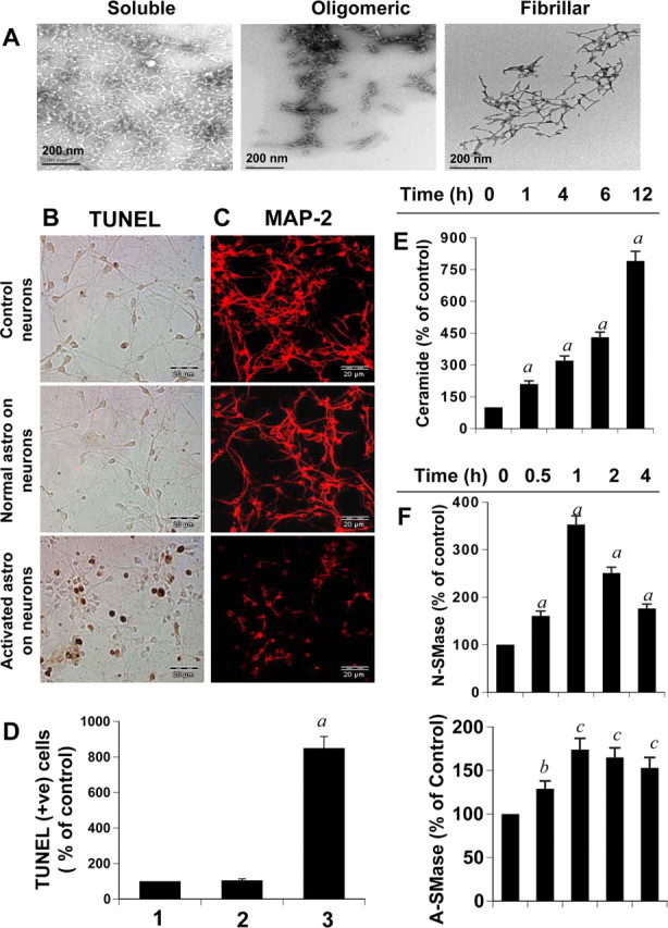
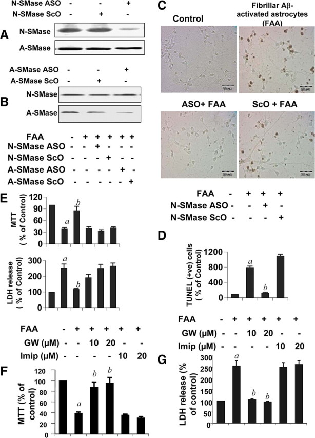
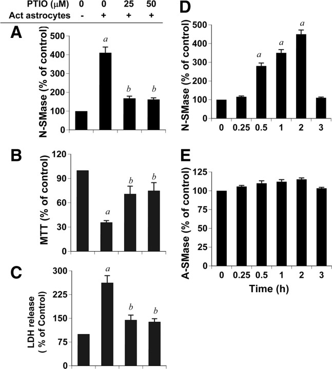
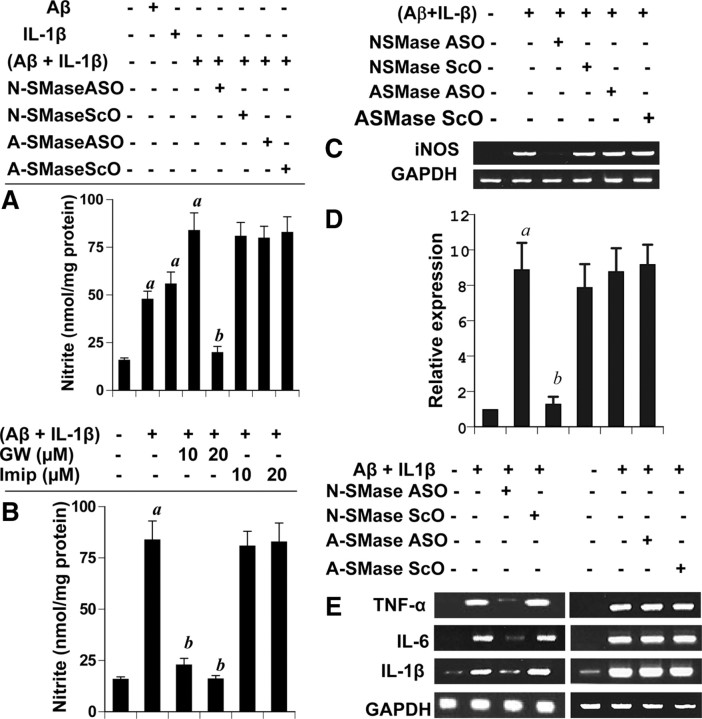
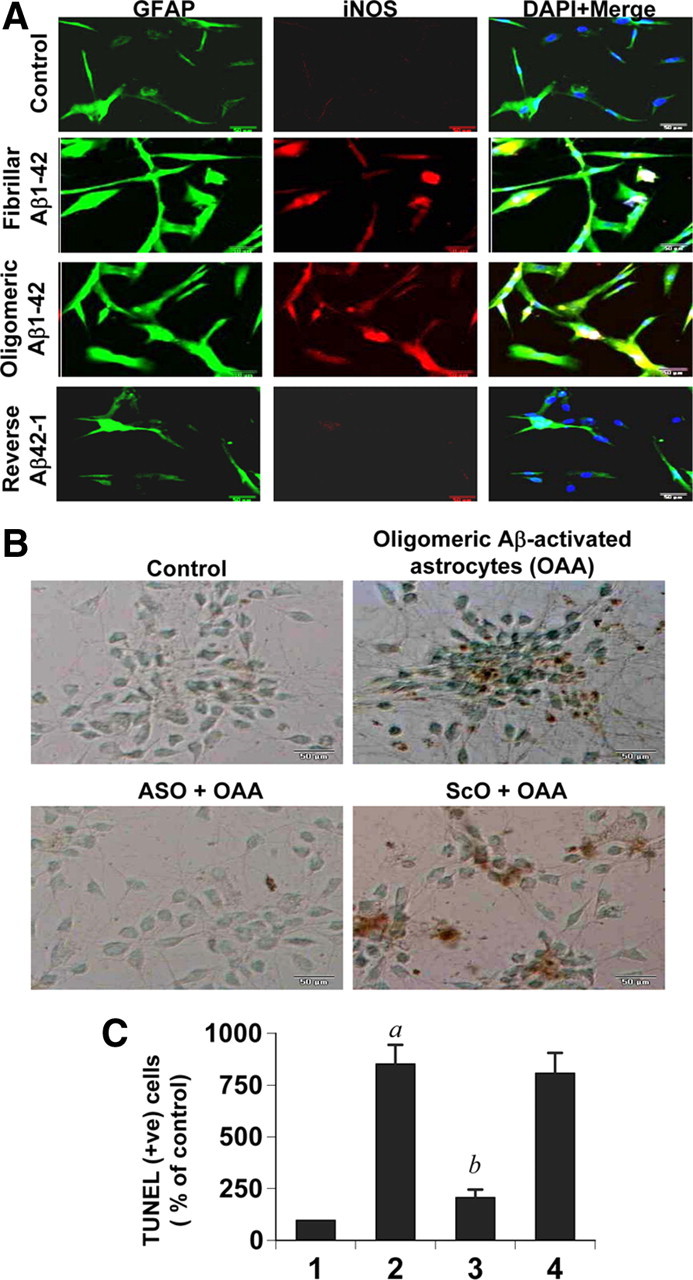
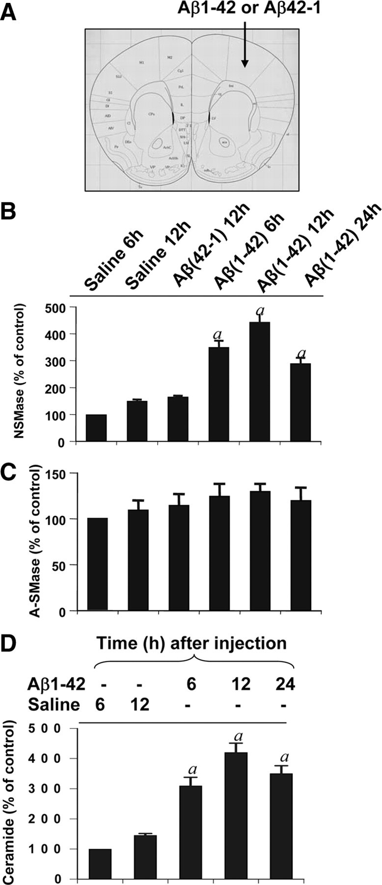
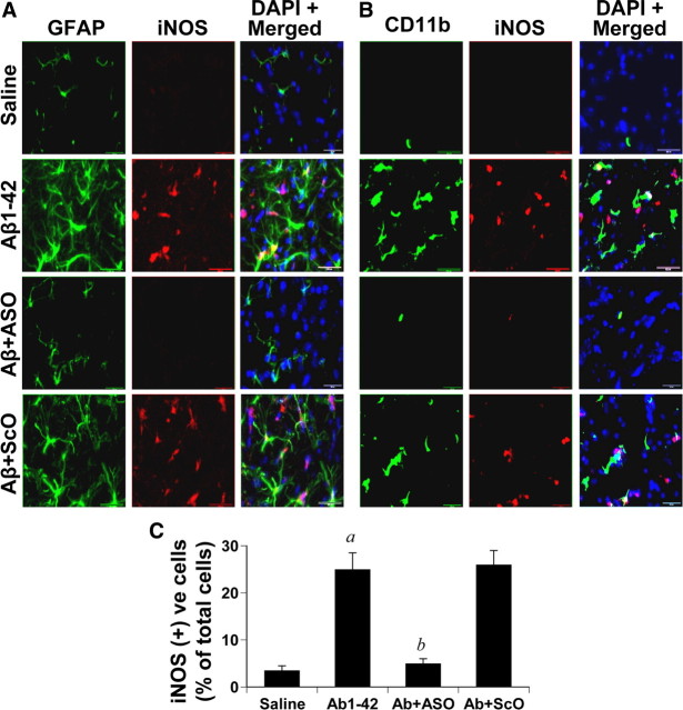
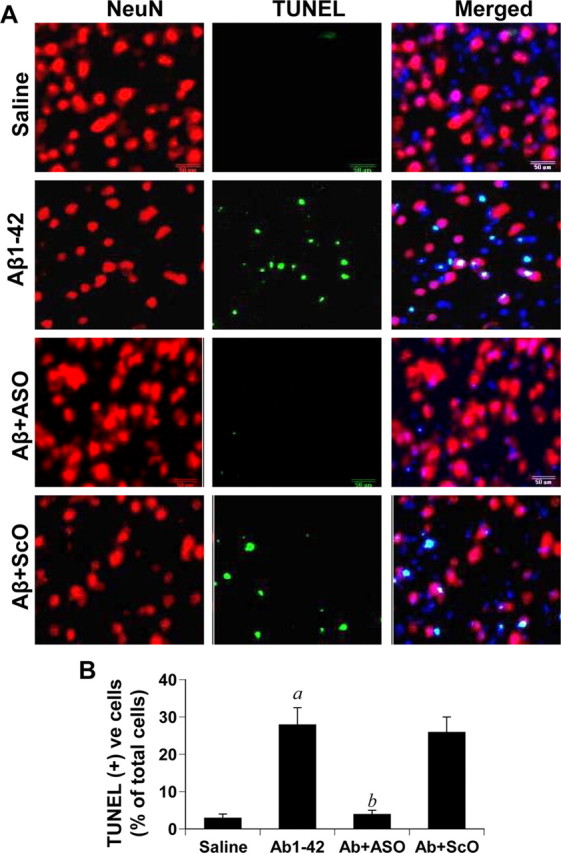
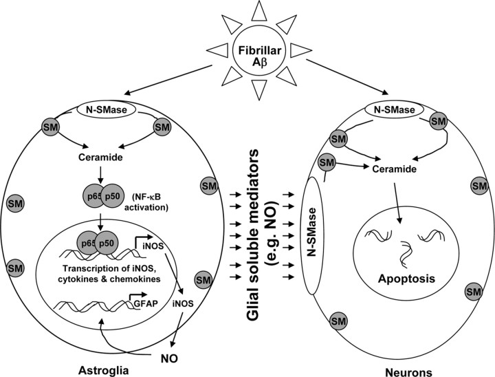
Glial activation plays an important role in the pathogenesis of various neurodegenerative disorders including Alzheimer's disease. However, molecular mechanisms by which activated glia could kill neurons are poorly understood. The present study underlines the importance of neutral sphingomyelinase (N-SMase) in mediating the damaging effect of fibrillar amyloid-β 1-42 (Aβ1-42) peptide-activated astroglia on neurons. In transwell experiments, soluble products released from activated primary human astroglia induced the activation of neutral sphingomyelinase (N-SMase), production of ceramide, and cell death in primary human neurons. Protection of neurons from cytotoxic effects of activated astroglia by antisense knockdown of N-SMase, but not acidic sphingomyelinase (A-SMase), suggests that soluble products released from activated astroglia kill neurons via N-SMase but not A-SMase. Next we examined the role of N-SMase in the activation of human astroglia. Interestingly, knockdown of N-SMase, but not A-SMase, by either antisense oligonucleotides or chemical inhibitor, prevented the induction of proinflammatory molecules [tumor necrosis factor-α, inducible nitric oxide synthase, interleukin-1β (IL-1β), and IL-6] and the activation of nuclear factor-κB in Aβ1-42-activated astroglia. Subsequently, fibrillar Aβ peptides also induced the activation of N-SMase and ceramide in vivo in mouse cortex. Most importantly, antisense knockdown of N-SMase, but not A-SMase, decreased the activation of astroglia and protected neurons from fibrillar Aβ toxicity in vivo in the cortex. Together, it is apparent that both the activation of astroglia by Aβ and that the cytotoxicity of activated astroglia on neurons depend on N-SMase.


Figures

Figure 1.
Figure 1.
Activated primary human astrocytes induce apoptosis and the activation of the N-SMase–ceramide pathway in primary human neurons in neuron–astrocyte transwell cultures. A, Morphology of oligomeric and fibrillar form of Aβ1-42 peptides was examined by transmission electron microscopy. B, Primary human astrocytes seeded in inserts were stimulated with a combination of 1 μm fibrillar Aβ and 10 ng/ml IL-1β in serum-free media. After 24 h, media were removed and inserts with activated astrocytes were placed on coverslips containing primary neurons for 6 h followed by TUNEL. C, After 18 h of treatment of neurons with activated astrocytes, cells were immunostained with MAP-2. D, TUNEL-positive cells were counted manually in four different images of each of three coverslips by three individuals blinded to the experiment. Values obtained from the control group (1) served as 100%, and data obtained from other two groups, i.e., the normal astrocyte–neuron group (2) and the activated astrocytes–neuron group (3) were calculated as a percentage of control accordingly. Results are the mean ± SD of three different experiments. ap < 0.001 versus control. Activated astrocytes were placed on human neurons and further incubated for different time intervals. E, Lipids were extracted from neurons at respective time points, and ceramide levels were determined. ap < 0.001 versus 0 h. F, Activities of N-SMase and A-SMase were assayed in total cell extract of neurons. Control group served as 100%, and data from other groups were expressed as a percentage of control. Results are the mean ± SD of three different experiments. ap < 0.001 versus 0 h; bp < 0.05 versus 0 h; cp < 0.001 versus 0 h.
Figure 2.
Figure 2.
Antisense or chemical knockdown of N-SMase, but not A-SMase, protects neurons from Aβ-activated primary human astrocytes in astrocyte–neuron transwell cultures. A, B, Primary human neurons were incubated with ASOs and ScOs against N-SMase (A) and A-SMase (B). After 48 h, neurons were analyzed for protein expression of N-SMase and A-SMase. C, Primary human astrocytes seeded in inserts were stimulated by the combination of Aβ and IL-1β as described above. After 24 h, media were removed, and inserts were washed and placed on neurons that were already pretreated for 40 h with 1 μm ASO or ScO against N-SMase and A-SMase. After 6 h of treatment with activated astrocytes, apoptotic events in neurons were detected by TUNEL. D, TUNEL-positive cells were counted manually in four different images of each of three coverslips by three individuals blinded to the experiment. E, After 18 h of stimulation, cell viability was examined by the metabolism of MTT and the release of LDH. Activated astrocytes were placed on neurons that were pretreated with different concentrations of GW4869 (GW) and imipramine. F, G, Cell viability was checked by MTT (F) and LDH (G). Values obtained from the control group served as 100%, and data obtained in other groups were calculated as a percentage of control accordingly. Results are the mean ± SD of three different experiments. ap < 0.001 versus control; bp < 0.001 versus FAA. FAA, Fibrillar Aβ+IL-1β-activated astrocytes.
Figure 3.
Figure 3.
Role of nitric oxide in activated astrocyte-induced cell death in neurons. Primary human astrocytes seeded in inserts were stimulated by the combination of Aβ and IL-1β as described above. After 24 h, media were removed and inserts were washed and placed on neurons that were pretreated with PTIO for 30 min. A, After 1 h, activity of N-SMase was measured in total cell extracts of neurons as described above. B, C, After 18 h, neuronal viability was examined by the metabolism of MTT (B) and the release of LDH (C). Values obtained from the control group served as 100%, and data obtained in other groups were calculated as a percentage of control accordingly. Results are the mean ± SD of three different experiments. ap < 0.001 versus control; bp < 0.001 versus activated astrocytes. D, E, Primary human neurons were treated with 25 μm DETA-NONOate (an NO donor), and at different time points of treatment, activities of N-SMase (D) and A-SMase (E) were monitored. ap < 0.001 versus control (0 h).
Figure 4.
Figure 4.
Antisense and chemical knockdown of N-SMase, but not A-SMase, blocks the expression of iNOS and proinflammatory cytokines in Aβ+IL-1β-activated primary human astrocytes. Cells pretreated with ASO and ScO against N-SMase and A-SMase for 40 h were stimulated with Aβ+IL-1β under the serum-free condition. A, After 24 h of stimulation, the concentration of nitrite was measured in supernatants by Griess reagent. B, Cells preincubated with different concentrations of GW4869 and imipramine for 30 min were stimulated as above and nitrite level was measured after 24 h of stimulation. Cells were treated with ASOs/ScOs for 40 h followed by stimulation with the combination of fibrillar Aβ and IL-1β as above. C, D, After 6 h, the expression of iNOS mRNA was analyzed by both semiquantitative RT-PCR (C) and quantitative real-time PCR (D). E, The expression of TNF-α, IL-6, and IL-1β mRNAs were analyzed by semiquantitative RT-PCR. Results are the mean ± SD of three different experiments. ap < 0.001 versus control; bp < 0.001 versus Aβ+IL-1β.
Figure 5.
Figure 5.
Oligomeric Aβ1-42-activated astrocytes induce apoptosis in neurons via N-SMase in neuron-astrocyte transwell cultures. A, Primary astrocytes were stimulated with fibrillar Aβ1-42, oligomeric Aβ1-42, and reverse peptide Aβ42-1 for 24 h followed by double labeling of GFAP and iNOS. B, Primary human astrocytes seeded in inserts were stimulated with a combination of 1 μm oligomeric Aβ1-42 and 10 ng/ml IL-1β in serum-free media. After 24 h, media were removed and inserts with activated astrocytes were placed on coverslips containing primary neurons that were already pretreated for 40 h with 1 μm ASO or ScO against N-SMase. After 6 h, neuronal apoptosis was monitored by TUNEL. C, TUNEL-positive cells were counted manually in four different images of each of three coverslips by three individuals blinded to the experiment. Values obtained from the control group (1) served as 100%, and data obtained from other three groups, i.e., the oligomeric Aβ-activated astrocytes (OAA) group (2), the ASO-OAA group (3), and the ScO-OAA group (4), were calculated as a percentage of control accordingly. Results are the mean ± SD of three different experiments. ap < 0.001 versus control; bp < 0.001 versus OAA.
Figure 6.
Figure 6.
Inhibition of N-SMase, but not A-SMase, suppresses the activation of NF-κB in activated primary human astrocytes. A, Cells treated with 1 μm ASO or ScO against N-SMase and A-SMase for 40 h in complete media were stimulated with Aβ+IL-1β under the serum-free condition. After 1 h of stimulation nuclear extracts were prepared and subjected to EMSA for the detection of NF-κB. B, C, In the other set of experiments, cells preincubated with different concentrations of GW4869 (B) and imipramine (C) for 30′ were stimulated with Aβ+IL-1β for 1 h followed by EMSA. The top and bottom arrows indicate the induced NF-κB band and the unbound probe, respectively. Results represent three independent experiments.
Figure 7.
Figure 7.
Fibrillar Aβ1-42 peptides induce the activation of N-SMase and the production of ceramide in vivo in the cortex of C57BL/6 mice. A, One microgram of either fibrillar Aβ1-42 or reverse Aβ42-1 peptides dissolved in saline was stereotaxically injected into the frontal cortex of C57BL/6 mice. Control mice received saline injection. B–D, At indicated time points, cortex was dissected out and divided into two halves: one half for measuring activities of N-SMase (B) and A-SMase (C) and the other for assaying ceramide (D). The control (saline, 6 h) group served as 100%, and data from other groups were expressed as a percentage of control. Results are the mean ± SD of five different mice (n = 5). ap < 0.05 versus saline (6 h).
Figure 8.
Figure 8.
Antisense knockdown of N-SMase attenuates the activation of astroglia and microglia and reduces the expression of iNOS in vivo in the cortex of C57BL/6 mice. One microgram of either ASO or ScO against N-SMase dissolved in 2 μl of saline was stereotaxically injected into the frontal cortex of C57BL/6 mice. After 24 h of microinjection, 1 μg of fibrillar Aβ1-42 in a 2 μl volume was again microinjected at the same site. A, B, After 6 h of Aβ microinjection, mice were perfused, and double immunofluorescence for GFAP (green) and iNOS (red) (A) and CD11b (green) and iNOS (red) (B) was performed. Five mice (n = 5) were used in each group. C, Cells positive for iNOS as well as total 4′,6′-diamidino-2-phenylindole-positive cells were counted in three cortical sections (two images per slide) of each of five different mice with an Olympus IX81 fluorescence microscope using the MicroSuite imaging software. Results are expressed as the number of iNOS-positive cells per 100 cells. ap < 0.001 versus saline; bp < 0.001 versus Aβ.
Figure 9.
Figure 9.
Antisense oligonucleotides against N-SMase protect neurons from fibrillar Aβ toxicity in vivo in the cortex of C57BL/6 mice. One microgram of either ASO or ScO against N-SMase dissolved in 2 μl of saline was stereotaxically injected into the frontal cortex. After 24 h of microinjection, 1 μg of fibrillar Aβ1-42 in a 2 μl volume was again microinjected at the same site. A, After 24 h of the final microinjection, mice were perfused and double immunofluorescence for NeuN (red), and TUNEL (green) was performed. Five mice (n = 5) were used in each group. B, TUNEL-positive cells as well as total 4′,6′-diamidino-2-phenylindole-positive cells were counted in three cortical sections (2 images per slide) of each of five different mice with an Olympus IX81 fluorescence microscope using the MicroSuite imaging software. Results are expressed as the number of TUNEL-positive cells per 100 cells. ap < 0.001 versus saline; bp < 0.001 versus Aβ.
Figure 10.
Figure 10.
Schematic presentation of the summary of our work. Fibrillar Aβ alone is capable of activating the N-SMase–ceramide pathway in neurons, which leads to apoptosis and cell death (Jana and Pahan, 2004a,b). In astroglia, fibrillar Aβ-mediated activation of the N-SMase–ceramide pathway is responsible for the activation of NF-κB and the expression of different proinflammatory soluble mediators including NO. Once NO is produced, it can induce the expression of GFAP in astrocytes leading to astrogliosis. On the other hand, these glial-derived soluble mediators (e.g., NO) also induce neuronal apoptosis via the N-SMase–ceramide pathway. It appears that multiple neurotoxic signaling pathways converge on N-SMase. SM, Sphingomyelin.

References

    1. Akiyama H, Barger S, Barnum S, Bradt B, Bauer J, Cole GM, Cooper NR, Eikelenboom P, Emmerling M, Fiebich BL, Finch CE, Frautschy S, Griffin WS, Hampel H, Hull M, Landreth G, Lue L, Mrak R, Mackenzie IR, McGeer PL, et al. Inflammation and Alzheimer's disease. Neurobiol Aging. 2000;21:383–421. - PMC - PubMed
    1. Auch CJ, Saha RN, Sheikh FG, Liu X, Jacobs BL, Pahan K. Role of protein kinase R in double-stranded RNA-induced expression of nitric oxide synthase in human astroglia. FEBS Lett. 2004;563:223–228. - PMC - PubMed
    1. Blackwell TS, Christman JW. The role of nuclear factor-kappa B in cytokine gene regulation. Am J Respir Cell Mol Biol. 1997;17:3–9. - PubMed
    1. Bodles AM, Barger SW. Secreted beta-amyloid precursor protein activates microglia via JNK and p38-MAPK. Neurobiol Aging. 2005;26:9–16. - PubMed
    1. Braak H, Braak E, Strothjohann M. Abnormally phosphorylated tau protein related to the formation of neurofibrillary tangles and neuropil threads in the cerebral cortex of sheep and goat. Neurosci Lett. 1994;171:1–4. - PubMed

Publication types

MeSH terms

LinkOut - more resources