. 2010 Dec;104(6):3424-32.
doi: 10.1152/jn.00540.2010. Epub 2010 Sep 22.

Reward magnitude coding in primate amygdala neurons

Affiliations

Reward magnitude coding in primate amygdala neurons

Maria A Bermudez et al. J Neurophysiol. 2010 Dec.

Abstract

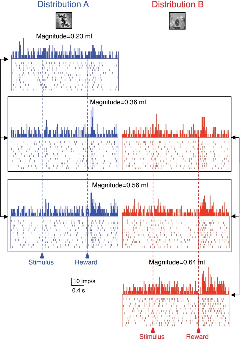
Animals assess the values of rewards to learn and choose the best possible outcomes. We studied how single neurons in the primate amygdala coded reward magnitude, an important variable determining the value of rewards. A single, Pavlovian-conditioned visual stimulus predicted fruit juice to be delivered with one of three equiprobable volumes (P = 1/3). A population of amygdala neurons showed increased activity after reward delivery, and almost one half of these responses covaried with reward magnitude in a monotonically increasing or decreasing fashion. A subset of the reward responding neurons were tested with two different probability distributions of reward magnitude; the reward responses in almost one half of them adapted to the predicted distribution and thus showed reference-dependent coding. These data suggest parametric reward value coding in the amygdala as a characteristic component of its function in reinforcement learning and economic decision making.


Figures

Fig. 1.
Fig. 1.
Task, licking, and recording sites. A: sequence of task events in the main experiment. The single stimulus predicted low, intermediate, or high magnitude of liquid reward with equal probability (P = 1/3) drawn semirandomly from distribution A. B: calibration of liquid reward magnitude as a function of solenoid valve opening time. The dashed lines indicate 4 typical values used for distributions A (main experiment) and B (additional for adaptation test). C: median lick durations during stimulus (●) and after reward onset (▴) for main distribution A (195 trials from 12 trial blocks). ○ and ▵, licking with the additional distribution B (adaptation test). The 95% CIs were (from left to right): stimulus A, 0.3, 0.1, and 0.1 (too small for error bars); stimulus B, 0.25, 0.25, and 0.1; rewards A, 83.3 (error bar), 0.4, and 0.5; rewards B, 95.4, 20.1, and 19.0. Trials were grouped according to the 2 distributions and the actual reward delivered (abscissa). D: histological reconstruction of recording sites in the amygdala for magnitude coding neurons in animal A, with approximate positions for animal B superimposed (total n = 56 neurons). CE, central nucleus; L, lateral nucleus; BL, basolateral nucleus; BM, basomedial nucleus.
Fig. 2.
Fig. 2.
Monotonic relationships of neuronal reward responses to variations in liquid magnitude. A: rasters and density functions of monotonically increasing responses from single amygdala neuron with reward magnitude. B: population density functions of averaged activity from all 46 positive monotonically coding neurons in both animals. C: monotonic decrease of neuronal reward responses with increasing liquid magnitude in single amygdala neuron (different neuron than shown in A). D: population density functions of averaged activity from all 10 inverse monotonically varying neurons. In A–D, red = 0.56 ml, green = 0.36 ml, blue = 0.23 ml, bin width = 10 ms, flat smoothing on 15 bins running, imp/s = impulses per second. E: increase in median normalized response strength (fractional change during 400-ms postreward period vs. 400-ms control period) as function of reward magnitude (±95% CIs; 46 neurons). Activity in each neuron was normalized to response to 0.56-ml reward, which itself consisted of a median increase of 251%. F: decrease in median normalized response strength as function of reward magnitude (10 neurons). Activity in each neuron was normalized to response to 0.23 ml reward, whose median increase was 284%.
Fig. 3.
Fig. 3.
Adaptive coding of reward magnitude in 1 amygdala neuron. The 3 reward magnitudes were delivered as elements of 2 different distributions (A: blue, left, B: red, right). Responses to the identical magnitudes varied depending on the distributions from which these magnitudes were drawn (0.36 and 0.56 ml, framed). Bin width = 10 ms.
Fig. 4.
Fig. 4.
Population display and analysis of adaptive reward magnitude coding in amygdala neurons. A: responses of the 14 adaptive amygdala neurons to delivery of 3 reward magnitudes as elements of 2 different distributions (A: blue, left, B: red, right). Bin width = 10 ms. B and C: median population responses to identical reward magnitudes in the adaptive neurons (B: 0.36 ml, n = 14 neurons; C: 0.56 ml, n = 13) delivered as elements of 2 different reward distributions (A vs. B). *P < 0.05; Mann-Whitney test. D and E: median responses of each neuron to reward magnitude = 0.36 ml (D) and magnitude = 0.56 ml (E) as elements of distribution A plotted against the same magnitudes as elements of distribution B. ●, significant response differences of the adaptive neurons between the 2 distributions in D (n = 14) and E (n = 13) (P < 0.05; Mann-Whitney test). ○, nonadaptive neurons (insignificantly different responses between distributions). Diagonal lines indicate identical responses with both distributions. Total n = 30 neurons in both D and E.
Fig. 5.
Fig. 5.
Absolute, nonadaptive coding of reward magnitude in 1 amygdala neuron. Three different reward magnitudes were delivered as elements of 2 different distributions (A: blue, left, B: red, right). Reward responses increased monotonically across all 4 magnitudes, irrespective of the distribution from which the magnitudes were drawn. Bin width = 10 ms.

Similar articles

Cited by

References

    1. Aggleton JP, Passingham RE. Stereotaxic surgery under X-ray guidance in the rhesus monkey, with special reference to the amygdala. Exp Brain Res 44: 271–276, 1981 - PubMed
    1. Apicella P, Ljungberg T, Scarnati E, Schultz W. Responses to reward in monkey dorsal and ventral striatum. Exp Brain Res 85: 491–500, 1991 - PubMed
    1. Baxter MG, Murray EA. The amygdala and reward. Nat Rev Neurosci 3: 563–573, 2002 - PubMed
    1. Baxter MG, Parker A, Lindner CCC, Izquierdo AD, Murray EA. Control of response selection by reinforcer value requires interaction of amygdala and orbital prefrontal cortex. J Neurosci 20: 4311–4319, 2000 - PMC - PubMed
    1. Bechara A, Damasio H, Damasio AR, Lee GP. Different contributions of the human amygdala and ventromedial prefrontal cortex to decision-making. J Neurosci 19: 5473–5481, 1999 - PMC - PubMed

Publication types

LinkOut - more resources