. 2010 Dec;104(6):3345-60.
doi: 10.1152/jn.00398.2010. Epub 2010 Sep 22.

Chronic dysfunction of astrocytic inwardly rectifying K+ channels specific to the neocortical epileptic focus after fluid percussion injury in the rat

Affiliations

Chronic dysfunction of astrocytic inwardly rectifying K+ channels specific to the neocortical epileptic focus after fluid percussion injury in the rat

Tessandra H Stewart et al. J Neurophysiol. 2010 Dec.

Abstract

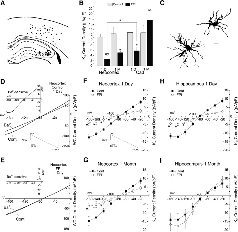
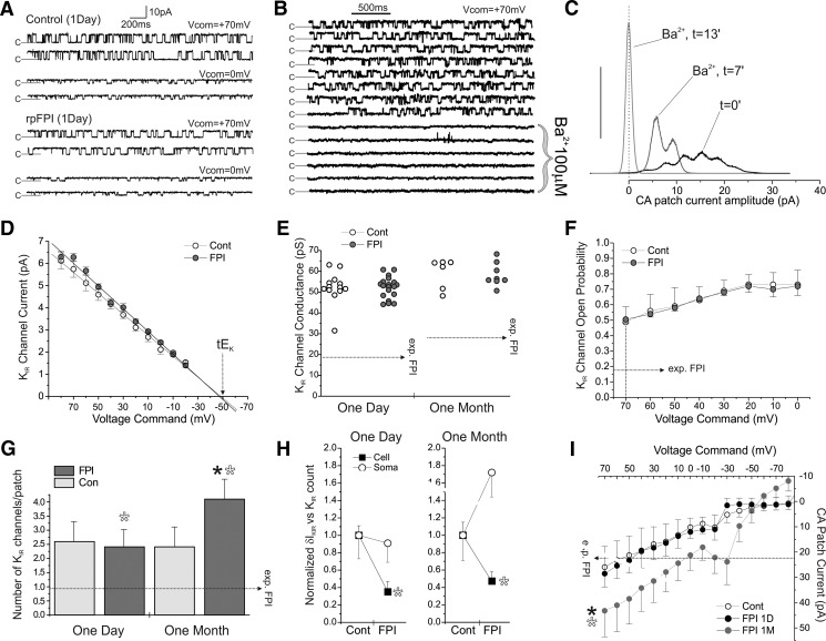
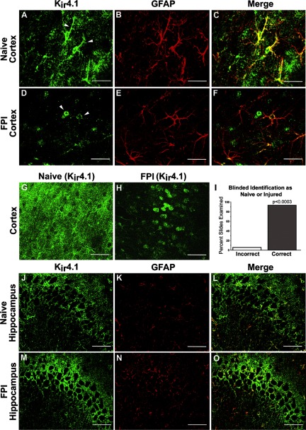
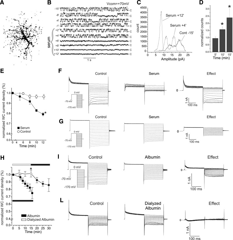
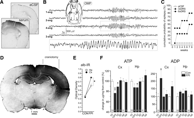
Astrocytic inwardly rectifying K(+) currents (I(KIR)) have an important role in extracellular K(+) homeostasis, which influences neuronal excitability, and serum extravasation has been linked to impaired K(IR)-mediated K(+) buffering and chronic hyperexcitability. Head injury induces acute impairment in astroglial membrane I(KIR) and impaired K(+) buffering in the rat hippocampus, but chronic spontaneous seizures appear in the perilesional neocortex--not the hippocampus--in the early weeks to months after injury. Thus we examined astrocytic K(IR) channel pathophysiology in both neocortex and hippocampus after rostral parasaggital fluid percussion injury (rpFPI). rpFPI induced greater acute serum extravasation and metabolic impairment in the perilesional neocortex than in the underlying hippocampus, and in situ whole cell recordings showed a greater acute loss of astrocytic I(KIR) in neocortex than hippocampus. I(KIR) loss persisted through 1 mo after injury only in the neocortical epileptic focus, but fully recovered in the hippocampus that did not generate chronic seizures. Neocortical cell-attached recordings showed no loss or an increase of I(KIR) in astrocytic somata. Confocal imaging showed depletion of KIR4.1 immunoreactivity especially in processes--not somata--of neocortical astrocytes, whereas hippocampal astrocytes appeared normal. In naïve animals, intracortical infusion of serum, devoid of coagulation-mediating thrombin activity, reproduces the effects of rpFPI both in vivo and at the cellular level. In vivo serum infusion induces partial seizures similar to those induced by rpFPI, whereas bath-applied serum, but not dialyzed albumin, rapidly silenced astrocytic K(IR) membrane currents in whole cell and cell-attached patch-clamp recordings in situ. Thus both acute impairment in astrocytic I(KIR) and chronic spontaneous seizures typical of rpFPI are reproduced by serum extravasation, whereas the chronic impairment in astroglial I(KIR) is specific to the neocortex that develops the epileptic focus.


Figures

Fig. 1.
Fig. 1.
Astrocytic KIR current loss after rostral parasaggital fluid percussion injury (rpFPI) persists chronically in the neocortex but not in the hippocampus. A: locations of the astrocytes sampled for in situ whole cell evaluation of KIR current. Patched cells from Bregma −0.5 to −3.5 mm are referred to a single anterior-posterior coordinate. B: group analysis of whole cell voltage-clamp recordings satisfying voltage-clamp quality control. Recordings were obtained from neocortical and CA3 astrocytes 1 day and 1 mo after FPI and in age-matched controls. KIR current density (δIKIR) is detected by its sensitivity to Ba2+ (40 μM) and was measured at −140 mV during voltage ramps from 0 to 170 mV. Ba2+-sensitive δIKIR is significantly decreased compared with age-matched controls in both neocortex and CA3 1 day after injury, but only in the neocortex 1 mo later. The acute loss in astrocytic δIKIR is more severe in the neocortex than in the hippocampus (*P < 0.025, **P < 0.0025). C: camera lucida images of confirmed isolated astrocytes filled with biocytin during patch-clamp recordings in brain slices obtained from FPI animals. Scale bar = 10 μm. D and E: representative whole cell current-voltage traces obtained with downward voltage ramps in neocortical astrocytes 1 day after rpFPI (E) and in age-matched controls (D), before (Cont) and after Ba2+ (40 μM) application, showing the acute loss in δIKIR. Top left insets in each panel show I-V plots of the Ba2+-sensitive δIKIR. F–I: cumulative I-V plots of the Ba2+-sensitive δIKIR measured during downward voltage ramps in the subset of astrocytes shown in B that were confirmed to be isolated. The δIKIR are plotted at 20 mV intervals from −160 to −20 mV. Ba2+-sensitive whole cell δIKIR was depressed 1 day after injury in both neocortex (F) and CA3 (H) and remained depressed at 1 mo only in neocortex (G), but not in CA3 where it recovered to preinjury values (I). Asterisks in F–I indicate statistical significance at the P < 0.01 level and, for clarity, are shown for voltage range from −160 to −120 mV only. Bottom right insets in D and F show the voltage ramp protocol.
Fig. 2.
Fig. 2.
Neocortical astrocytic somata present no change in KIR current at 1 day and a compensatory increase at 1 mo after rpFPI. Cell-attached recordings from astrocytic somata do not show loss of IKIR, suggesting the IKIR loss observed in whole cell is from the impairment of astrocytes' processes. A: channel gating (c = closed) during cell-attached recordings from control and rpFPI neocortical astrocytic somata at Vcom of +70 and 0 mV. B: segments of consecutive sweeps obtained over 15 min from a cell-attached patch containing 1 active KIR channel before and during Ba2+ (100 μM) application through the pipette. Channel opening is potently blocked by Ba2+ as expected for KIR channels. C: all-points histograms of KIR channel activity during application of 100 μM Ba2+ through the pipette to a patch bearing multiple channels. Peaks at t = 0 indicate opening of multiple channels before Ba2+ application. Peaks are left-shifted and consolidated over time, indicating Ba2+ blockade of all channels on the patch. At t = 13 min, all channels are permanently closed. Y scale in arbitrary units. D: current-voltage plots for control and rpFPI channels recorded in somatic cell-attached patches show the same reversal potential (Vcom ≈ −48mV) consistent with a theoretical EK (tEK) in cell-attached of ≈ −22 mV, assuming cell Vm ≈ −70 mV, [K+]i = 66 mM, and [K+]pipette = 140 mM. E: slope conductance of cell-attached KIR channels from control and FPI rats 1 day and 1 mo after FPI. No injury-induced changes in channel conductance are observed. F: voltage dependence of Po in channels from control and 1 day FPI patches. No injury-induced changes in channel Po are observed. G: numbers of KIR channels found in cell-attached patches obtained 1 day and 1 mo after injury. No changes are observed 1 day after rpFPI, when the whole cell δIKIR deficit is most severe, whereas an increase in number of KIR channels is observed 1 mo after injury. Filled asterisk indicates statistical significant difference of rpFPI (1 mo) compared with a pooled control incorporating all other groups (P = 0.027). Hollow asterisks indicate statistical significance of the 2-way ANOVA test shown in H. H: summary of the 2-way ANOVA indicating a statistically significant difference between the effects of rpFPI on whole cell δIKIR (Cell) and on cell-attached KIR channel count (Soma). Cell values are normalized to the mean of the control groups. Asterisks indicate statistical significance: 1 day, P = 0.025; 1 mo, P = 0.002. I: I-V plot of mean cell-attached patch currents (corrected for leak current through the gigaseal) recorded during applied potentials from +70 to −70 mV in control and FPI at 1 day and 1 mo after injury. Currents measured 1 day after FPI are similar to control but are increased at 1 mo after FPI. Filled asterisk indicates P < 0.05 compared with controls. Empty asterisk indicates the significant difference (treatment × technique interaction) in the effect of rpFPI on somatic cell-attached vs. whole cell inward currents measured in the same cells (P = 0.003). Horizontal dotted arrows (exp. FPI) in E–I indicate the expected changes required to fully account for the whole cell KIR deficit after FPI.
Fig. 3.
Fig. 3.
FPI induces chronic KIR4.1 mislocalization in cortical but not hippocampal astrocytes. Confocal microscopic images of KIR4.1 (green) and glial fibrillary acid protein (GFAP; red) immunofluorescence were obtained from regions of neocortex (A–H) and CA3 hippocampus (J–O), corresponding to the regions from which astroglial patch-clamp recordings were obtained (see Fig. 1A). Controls are shown in A–C, G, and J–L. Sections obtained at 1 mo after FPI are shown in D–F, H, and M–O. G, H, and all right-most images are merged images showing both KIR4.1 and GFAP immunoreactivity. A–C: naïve control cortex. KIR4.1 was expressed prominently in GFAP-positive astrocytic processes (arrowheads), as well as in astrocytic cell bodies. D–F: perilesional rpFPI cortex 1 mo after injury. Compared with controls, KIR4.1 immunoreactivity in injured cortical astrocytes (D–F) appears more prominent in swollen-appearing astrocytic cell bodies (arrowheads), whereas KIR4.1 immunoreactivity in GFAP-positive processes was markedly reduced. G and H: lower-magnification images of naïve (G) and injured cortex (H) similarly show that FPI was associated with an apparent loss of KIR4.1 immunoreactivity in finely ramifying processes. I: results of a histological analysis carried out under double-blind conditions that confirmed that the patterns of KIR4.1 expression in the somata and processes of astrocytes (D, F, and H) were distinct in naive and perilesional FPI neocortices. P = exact binomial probability. J–O: in contrast to the injury-induced Kir4.1 mislocalization observed in neocortex, the expression pattern of Kir4.1 in the CA3 region of hippocampus of injured rats 30 days after injury (M–O) was comparable to controls (J–L). Scale bars: C, F, L, and O: 50 μm; G and H: 100 μm.
Fig. 4.
Fig. 4.
Serum, but not dialyzed albumin, induces acute loss of neocortical astrocyte KIR channel activity under conditions of blocked neuronal and synaptic activity. A: brightest point projection of an astrocyte with complex electrophysiological profile filled in situ with rhodamine dextran after cell-attached recordings. B: the activity of a single KIR channel, recorded from a neocortical astrocytic soma in a brain slice, is shown in sweeps obtained at a command potential of +70 mV before (t = −15 to 0 min) and during (t = 0–2 min) exposure to threefold diluted rat serum (DRS). Channel activity, stable over the course of 15 min before DRS application, ceased abruptly after 4 min in DRS. C: all-points histograms obtained at a command potential of +70 mV from a somatic cell-attached patch from another neocortical astrocyte, in situ, displayed numerous stable conductance states before DRS application, and just 1 (closed) afterward. The increase in peak amplitude and decrease in the width of the histogram during DRS exposure indicate a progressive shut-down of the channels in the patch. After 12 min of DRS exposure, all channel activity has ceased, resulting in an all-points histogram with a single, narrow high-amplitude peak. D: group analysis of peak amplitudes in all-points histograms obtained in cell-attached recordings after 5–15 min of DRS exposure normalized to preserum control amplitudes. Asterisks indicate P < 0.05. E: group analysis of the effect of exposure of neocortical astrocytes in situ to threefold DRS. DRS induces a time-dependent loss of whole cell δIKIR during upward voltage ramps. Plot shows time course of δIKIR measured at −140 mV and normalized to pretreatment values. Slices were treated with artificial cerebrospinal fluid (ACSF; control) or threefold DRS (serum) both with kynurenic acid (KYNA; 1 mM) and TTX (1 μM) to prevent epileptiform activity. F and G: whole cell current response to descending voltage steps in 2 in situ astrocytes before (control) and after (serum) ∼12 min of exposure to DRS. KIR current was lost (effect) in both complex (F) and noncomplex (G) astrocytes. Inset: voltage-clamp commands. H: group analysis of the effect of exposure to 0.4 mM whole bovine serum albumin (BSA; 99% purity; filled squares), or 0.4 mM dialyzed albumin (gray diamonds), in KYNA (1 mM). Whole BSA induces a serum like time-dependent loss of whole cell δIKIR during upward voltage ramps in naïve neocortical astrocytes that is not reproduced by dialyzed albumin (asterisk indicates P < 0.05). Note the positive effect of whole albumin applied to the same cells after the ineffective exposure to dialyzed albumin. Plot shows time course of δIKIR measured at −140 mV and normalized to pretreatment values. Black and gray bars indicate the exposure to whole albumin or dialyzed albumin, respectively. I: whole cell current response to descending voltage steps in an in situ astrocyte before (control) and after (albumin) 15 min of exposure to 99% pure albumin in KYNA (1 mM). KIR current was lost (effect). Inset: voltage-clamp protocol. Note the abolished KIR current in the bottom panel. J: whole cell current recordings during voltage clamp (VC) protocols identical to that used in G in an in situ astrocyte before (control) and after (dialyzed albumin) 15 min of exposure to dialyzed albumin in KYNA (1 mM). KIR current was not affected (effect).
Fig. 5.
Fig. 5.
Serum infusion in vivo induces chronic recurrent spontaneous seizures and demonstration of a greater severity of injury suffered by the neocortex than the underlying hippocampus. A: albumin immunoreactivity marks the area affected by 6 days of in vivo infusion of either threefold autologous DRS or ACSF into the neocortical parenchyma of naïve rats, through a glass micropipette. Infusion of DRS results in prominent albumin immunoreactivity throughout much of the surrounding neocortex and dorsal hippocampus (serum), whereas there was no detectable albumin extravasation after infusion of ACSF. B: a representative grade 2 focal seizure recorded 2 wk after infusion of threefold DRS. Dotted box delimits the portion of ECoG shown at higher temporal resolution. The numbers next to each ECoG trace indicate the electrode (inset) by which the trace was recorded and the average reference (avg). Inset: schematic of the rat skull and 5-electrode epidural ECoG montage (1–5) used to detect serum induced neocortical seizures. Empty circle represents the site of insertion of the glass micropipette for intraparenchymal infusion of DRS or ACSF by osmotic minipump (OMP) in vivo. C: time course of epileptogenesis after intracortical infusion of ACSF, threefold DRS or sixfold DRS for 6 days. Whereas seizures were not observed in any ACSF infused animal, those receiving DRS developed epilepsy in a dose-dependent fashion. D: albumin immunoreactivity (alb-IR) marks extensive blood–brain barrier compromise and serum extravasation in the neocortex and hippocampus 6 h after rpFPI. Craniotomy indicates site of rpFPI delivery. E: densitometric analysis of the effect of injury on alb-IR in neocortex (Cx) and hippocampus (Hp). The FPI-induced increase in albIR was ∼50% greater in neocortex than hippocampus (P < 0.05, Wilcoxon). C: changes in tissue ATP and ADP content in the ipsilateral rostral parietal neocortex (Cx) and hippocampus (Hp) over time after injury. The neocortex suffers greater disruption of energy metabolism than the hippocampus. Numbers at the base of each column indicate group size. Asterisks in E and F indicate statistical significance (*P < 0.05, **P < 0.005) compared with age-matched controls.

Similar articles

Cited by

References

    1. Abrahám H, Losonczy A, Czéh G, Lázár G. Rapid activation of microglial cells by hypoxia, kainic acid, and potassium ions in slice preparations of the rat hippocampus. Brain Res 906: 115–126, 2001 - PubMed
    1. Albensi BC, Janigro D. Traumatic brain injury and its effects on synaptic plasticity. Brain Inj 17: 653–663, 2003 - PubMed
    1. Alexander JS, Patton WF, Christman BW, Cuiper LL, Haselton FR. Platelet-derived lysophosphatidic acid decreases endothelial permeability in vitro Am J Physiol Heart Circ Physiol 274: H115–H122, 1998 - PubMed
    1. Annegers JF, Hauser WA, Coan SP, Rocca WA. A population-based study of seizures after traumatic brain injuries. N Engl J Med 338: 20–24, 1998 - PubMed
    1. Ballanyi K, Grafe P, ten Bruggencate G. Ion activities and potassium uptake mechanisms of glial cells in guinea-pig olfactory cortex slices. J Physiol 382: 159–174, 1987 - PMC - PubMed

Publication types

MeSH terms

LinkOut - more resources