

Seizure identification in the ICU using quantitative EEG displays
- PMID: 20861452
- PMCID: PMC2974462
- DOI: 10.1212/WNL.0b013e3181f9619e
Seizure identification in the ICU using quantitative EEG displays
Abstract
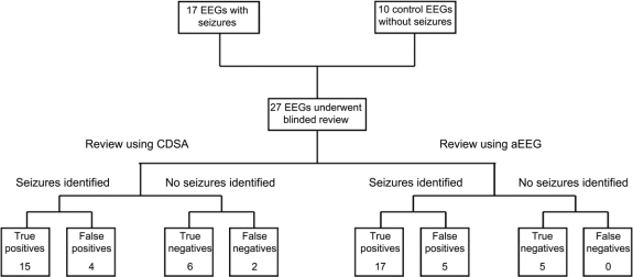
Objective: To evaluate the diagnostic accuracy of 2 quantitative EEG display tools, color density spectral array (CDSA) and amplitude-integrated EEG (aEEG), for seizure identification in the intensive care unit (ICU).
Methods: A set of 27 continuous EEG recordings performed in pediatric ICU patients was transformed into 8-channel CDSA and aEEG displays. Three neurophysiologists underwent 2 hours of training to identify seizures using these techniques. They were then individually presented with a series of CDSA and aEEG displays, blinded to the raw EEG, and asked to mark any events suspected to be seizures. Their performance was compared to seizures identified on the underlying conventional EEG.
Results: The 27 EEG recordings contained 553 discrete seizures over 487 hours. The median sensitivity for seizure identification across all recordings was 83.3% using CDSA and 81.5% using aEEG. However, among individual recordings, the sensitivity ranged from 0% to 100%. Factors reducing the sensitivity included low-amplitude, short, and focal seizures. False-positive rates were generally very low, with misidentified seizures occurring once every 17-20 hours.
Conclusions: Both CDSA and aEEG demonstrate acceptable sensitivity and false-positive rates for seizure identification among critically ill children. Accuracy of these tools would likely improve during clinical use, when findings can be correlated in real-time with the underlying raw EEG. In the hands of neurophysiologists, CDSA and aEEG displays represent useful screening tools for seizures during continuous EEG monitoring in the ICU. The suitability of these tools for bedside use by ICU nurses and physicians requires further study.
Figures


Similar articles
-
Detection of seizure patterns with multichannel amplitude-integrated EEG and the color density spectral array in the adult neurology intensive care unit.Medicine (Baltimore). 2018 Sep;97(38):e12514. doi: 10.1097/MD.0000000000012514. Medicine (Baltimore). 2018. PMID: 30235767 Free PMC article. Clinical Trial.
-
Seizure Detection by Critical Care Providers Using Amplitude-Integrated Electroencephalography and Color Density Spectral Array in Pediatric Cardiac Arrest Patients.Pediatr Crit Care Med. 2017 Apr;18(4):363-369. doi: 10.1097/PCC.0000000000001099. Pediatr Crit Care Med. 2017. PMID: 28234810 Free PMC article. Clinical Trial.
-
Non-expert use of quantitative EEG displays for seizure identification in the adult neuro-intensive care unit.Epilepsy Res. 2015 Jan;109:48-56. doi: 10.1016/j.eplepsyres.2014.10.013. Epub 2014 Oct 28. Epilepsy Res. 2015. PMID: 25524842
-
A Comparison Of Conventional Electroencephalography With Amplitude-Integrated EEG In Detection Of Neonatal Seizures.Med Devices (Auckl). 2019 Dec 10;12:489-496. doi: 10.2147/MDER.S214662. eCollection 2019. Med Devices (Auckl). 2019. PMID: 31849541 Free PMC article. Review.
-
Amplitude-integrated electroencephalography for seizure detection in newborn infants.Semin Fetal Neonatal Med. 2018 Jun;23(3):175-182. doi: 10.1016/j.siny.2018.02.003. Epub 2018 Feb 13. Semin Fetal Neonatal Med. 2018. PMID: 29472139 Review.
Cited by
-
Predicting outcome in patients with moderate to severe traumatic brain injury using electroencephalography.Crit Care. 2019 Dec 11;23(1):401. doi: 10.1186/s13054-019-2656-6. Crit Care. 2019. PMID: 31829226 Free PMC article.
-
Adult Critical Care Electroencephalography Monitoring for Seizures: A Narrative Review.Front Neurol. 2022 Jul 15;13:951286. doi: 10.3389/fneur.2022.951286. eCollection 2022. Front Neurol. 2022. PMID: 35911927 Free PMC article. Review.
-
Diagnostic accuracy between readers for identifying electrographic seizures in critically ill adults.Epilepsia Open. 2017 Jan 3;2(1):67-75. doi: 10.1002/epi4.12034. eCollection 2017 Mar. Epilepsia Open. 2017. PMID: 29750214 Free PMC article.
-
Early ketamine to treat refractory status epilepticus.Neurocrit Care. 2012 Apr;16(2):299-305. doi: 10.1007/s12028-011-9668-7. Neurocrit Care. 2012. PMID: 22237581
-
Density spectral array for seizure identification in critically ill children.J Clin Neurophysiol. 2013 Aug;30(4):371-5. doi: 10.1097/WNP.0b013e31829de01c. J Clin Neurophysiol. 2013. PMID: 23912575 Free PMC article.
References
-
- Friedman D, Claassen J, Hirsch LJ. Continuous electroencephalogram monitoring in the intensive care unit. Anesth Analg 2009;109:506–523. - PubMed
-
- Jirsch J, Hirsch LJ. Nonconvulsive seizures: developing a rational approach to the diagnosis and management in the critically ill population. Clin Neurophysiol 2007;118:1660–1670. - PubMed
-
- Jette N, Claassen J, Emerson RG, Hirsch LJ. Frequency and predictors of nonconvulsive seizures during continuous electroencephalographic monitoring in critically ill children. Arch Neurol 2006;63:1750–1755. - PubMed
-
- Claassen J, Mayer SA, Kowalski RG, Emerson RG, Hirsch LJ. Detection of electrographic seizures with continuous EEG monitoring in critically ill patients. Neurology 2004;62:1743–1748. - PubMed
Publication types
MeSH terms
Grants and funding
LinkOut - more resources
Full Text Sources
Other Literature Sources
Medical