

Tight junction-associated signaling pathways modulate cell proliferation in uveal melanoma
- PMID: 20861479
- PMCID: PMC3053300
- DOI: 10.1167/iovs.10-5746
Tight junction-associated signaling pathways modulate cell proliferation in uveal melanoma
Abstract
Purpose: To investigate the role of tight junction (TJ)-associated signaling pathways in the proliferation of uveal melanoma.
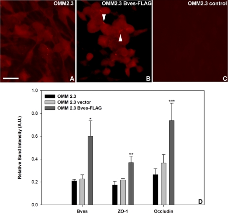
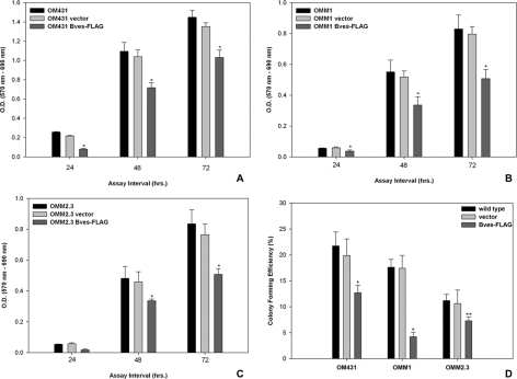
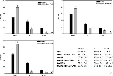
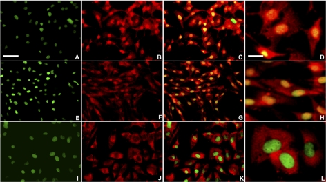
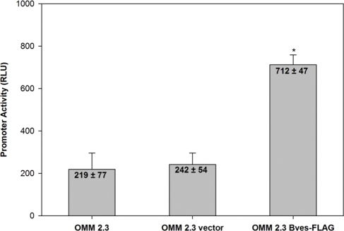
Methods: Human uveal melanoma cell lines overexpressing the TJ molecule blood vessel epicardial substance (Bves) were generated. The effects of Bves overexpression on TJ protein expression, cell proliferation, and cell cycle distribution were quantified. In addition, localization and transcription activity of the TJ-associated protein ZO-1-associated nucleic acid binding protein (ZONAB) were evaluated using immunofluorescence and bioluminescence reporter assays to study the involvement of Bves signaling in cell proliferation-associated pathways.
Results: Bves overexpression in uveal melanoma cell lines resulted in increased expression of the TJ proteins occludin and ZO-1, reduced cell proliferation, and increased sequestration of ZONAB at TJs and reduced ZONAB transcriptional activity.
Conclusions: TJ proteins are present in uveal melanoma, and TJ-associated signaling pathways modulate cell signaling pathways relevant to proliferation in uveal melanoma.
Figures
Similar articles
-
Inhibition of RhoA signaling with increased Bves in trabecular meshwork cells.Invest Ophthalmol Vis Sci. 2010 Jan;51(1):223-30. doi: 10.1167/iovs.09-3539. Epub 2009 Jul 23. Invest Ophthalmol Vis Sci. 2010. PMID: 19628742 Free PMC article.
-
Bves modulates tight junction associated signaling.PLoS One. 2011 Jan 20;6(1):e14563. doi: 10.1371/journal.pone.0014563. PLoS One. 2011. PMID: 21283798 Free PMC article.
-
Regulation of PCNA and cyclin D1 expression and epithelial morphogenesis by the ZO-1-regulated transcription factor ZONAB/DbpA.Mol Cell Biol. 2006 Mar;26(6):2387-98. doi: 10.1128/MCB.26.6.2387-2398.2006. Mol Cell Biol. 2006. PMID: 16508013 Free PMC article.
-
Phase separation as a therapeutic target in tight junction-associated human diseases.Acta Pharmacol Sin. 2020 Oct;41(10):1310-1313. doi: 10.1038/s41401-020-0470-y. Epub 2020 Jul 21. Acta Pharmacol Sin. 2020. PMID: 32694756 Free PMC article. Review.
-
Structural and signalling molecules come together at tight junctions.Curr Opin Cell Biol. 1999 Oct;11(5):628-33. doi: 10.1016/s0955-0674(99)00016-2. Curr Opin Cell Biol. 1999. PMID: 10508648 Review.
Cited by
-
Tight Junctions in Cell Proliferation.Int J Mol Sci. 2019 Nov 27;20(23):5972. doi: 10.3390/ijms20235972. Int J Mol Sci. 2019. PMID: 31783547 Free PMC article. Review.
-
Cold Shock Proteins Mediate GN with Mesangioproliferation.J Am Soc Nephrol. 2016 Dec;27(12):3678-3689. doi: 10.1681/ASN.2015121367. Epub 2016 May 5. J Am Soc Nephrol. 2016. PMID: 27151923 Free PMC article.
-
Reduced Popdc3 expression correlates with high risk and poor survival in patients with gastric cancer.World J Gastroenterol. 2012 May 21;18(19):2423-9. doi: 10.3748/wjg.v18.i19.2423. World J Gastroenterol. 2012. PMID: 22654436 Free PMC article.
-
Correlation of histopathologic characteristics to protein expression and function in malignant melanoma.PLoS One. 2017 Apr 26;12(4):e0176167. doi: 10.1371/journal.pone.0176167. eCollection 2017. PLoS One. 2017. PMID: 28445515 Free PMC article.
-
The Popeye domain containing protein family--A novel class of cAMP effectors with important functions in multiple tissues.Prog Biophys Mol Biol. 2016 Jan;120(1-3):28-36. doi: 10.1016/j.pbiomolbio.2016.01.001. Epub 2016 Jan 7. Prog Biophys Mol Biol. 2016. PMID: 26772438 Free PMC article. Review.
References
-
- Singh AD, Kivela T. The collaborative ocular melanoma study. Ophthalmol Clin North Am. 2005;18:129–142, ix - PubMed
-
- Gamel JW, McLean IW. Quantitative analysis of the Callender classification of uveal melanoma cells. Arch Ophthalmol. 1977;95:686–691 - PubMed
-
- Folberg R, Rummelt V, Parys-Van Ginderdeuren R, et al. The prognostic value of tumor blood vessel morphology in primary uveal melanoma. Ophthalmology. 1993;100:1389–1398 - PubMed
-
- Folberg R, Pe'er J, Gruman LM, et al. The morphologic characteristics of tumor blood vessels as a marker of tumor progression in primary human uveal melanoma: a matched case-control study. Hum Pathol. 1992;23:1298–1305 - PubMed
Publication types
MeSH terms
Substances
Grants and funding
LinkOut - more resources
Full Text Sources
Medical
Miscellaneous
