

Immunological control of adult neural stem cells
- PMID: 20861925
- PMCID: PMC2946325
Immunological control of adult neural stem cells
Abstract
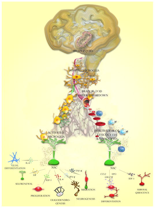
Adult neurogenesis occurs only in discrete regions of adult central nervous system: the subventricular zone and the subgranular zone. These areas are populated by adult neural stem cells (aNSC) that are regulated by a number of molecules and signaling pathways, which control their cell fate choices, survival and proliferation rates. For a long time, it was believed that the immune system did not exert any control on neural proliferative niches. However, it has been observed that many pathological and inflammatory conditions significantly affect NSC niches. Even more, increasing evidence indicates that chemokines and cytokines play an important role in regulating proliferation, cell fate choices, migration and survival of NSCs under physiological conditions. Hence, the immune system is emerging is an important regulator of neurogenic niches in the adult brain, which may have clinical relevance in several brain diseases.
Figures

Similar articles
-
Immune system modulates the function of adult neural stem cells.Curr Immunol Rev. 2010 Aug 1;6(3):167-173. doi: 10.2174/157339510791823772. Curr Immunol Rev. 2010. PMID: 21037937 Free PMC article.
-
Neuro-immune interactions in the postnatal ventricular-subventricular zone.J Stem Cells. 2014;9(1):53-64. J Stem Cells. 2014. PMID: 25158089
-
Grafted Subventricular Zone Neural Stem Cells Display Robust Engraftment and Similar Differentiation Properties and Form New Neurogenic Niches in the Young and Aged Hippocampus.Stem Cells Transl Med. 2016 Sep;5(9):1204-15. doi: 10.5966/sctm.2015-0270. Epub 2016 May 18. Stem Cells Transl Med. 2016. PMID: 27194744 Free PMC article.
-
Immunological regulation of neurogenic niches in the adult brain.Neuroscience. 2012 Dec 13;226:270-81. doi: 10.1016/j.neuroscience.2012.08.053. Epub 2012 Sep 15. Neuroscience. 2012. PMID: 22986164 Free PMC article. Review.
-
Adult neural stem cells, neurogenic niches, and cellular therapy.Stem Cell Rev. 2006;2(3):213-9. doi: 10.1007/s12015-006-0049-0. Stem Cell Rev. 2006. PMID: 17625257 Review.
Cited by
-
Ethical implications in the use of embryonic and adult neural stem cells.Stem Cells Int. 2012;2012:470949. doi: 10.1155/2012/470949. Epub 2012 Sep 10. Stem Cells Int. 2012. PMID: 22997522 Free PMC article.
-
Neuroimmunomodulation in unipolar depression: a focus on chronobiology and chronotherapeutics.J Neural Transm (Vienna). 2012 Oct;119(10):1147-66. doi: 10.1007/s00702-012-0819-6. Epub 2012 Jun 1. J Neural Transm (Vienna). 2012. PMID: 22653515 Review.
-
Role of neuroinflammation in adult neurogenesis and Alzheimer disease: therapeutic approaches.Mediators Inflamm. 2013;2013:260925. doi: 10.1155/2013/260925. Epub 2013 Apr 3. Mediators Inflamm. 2013. PMID: 23690659 Free PMC article. Review.
-
Mechanisms and Functional Significance of Stroke-Induced Neurogenesis.Front Neurosci. 2015 Dec 8;9:458. doi: 10.3389/fnins.2015.00458. eCollection 2015. Front Neurosci. 2015. PMID: 26696816 Free PMC article. Review.
-
Transplantation of adult monkey neural stem cells into a contusion spinal cord injury model in rhesus macaque monkeys.Cell J. 2014 Summer;16(2):117-130. Epub 2014 May 25. Cell J. 2014. PMID: 24567941 Free PMC article.
References
-
- Altman J. Postnatal neurogenesis and the problem of neural plasticity. In: Himwich WA, editor. Developmental neurobiology. C.C.Thomas; Springfield: 1970. pp. 197–237.
-
- Alvarez-Buylla A, Nottebohm F. Seasonal and species differences in the production of long projection neurons in adult birds. Neuroscience. 1989;15:962.
-
- Alvarez-Buylla A, Garcia-Verdugo JM, Tramontin AD. A unified hypothesis on the lineage of neural stem cells. Nat Rev Neurosci. 2001;2. 2(4. 4):287–93. - PubMed
Publication types
MeSH terms
Grants and funding
LinkOut - more resources
Full Text Sources