. 2010 Sep 17;5(9):e12837.
doi: 10.1371/journal.pone.0012837.

Bone is not essential for osteoclast activation

Affiliations

Bone is not essential for osteoclast activation

Karen Fuller et al. PLoS One. .

Abstract

Background: The mechanism whereby bone activates resorptive behavior in osteoclasts, the cells that resorb bone, is unknown. It is known that α(v)β(3) ligands are important, because blockade of α(v)β(3) receptor signaling inhibits bone resorption, but this might be through inhibition of adhesion or migration rather than resorption itself. Nor is it known whether α(v)β(3) ligands are sufficient for resorption the consensus is that bone mineral is essential for the recognition of bone as the substrate appropriate for resorption.

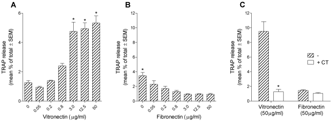
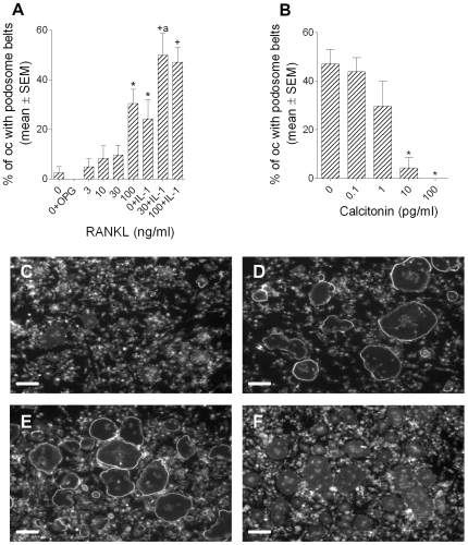
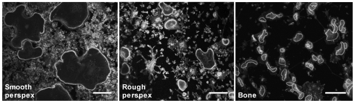
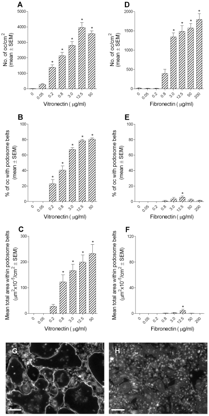
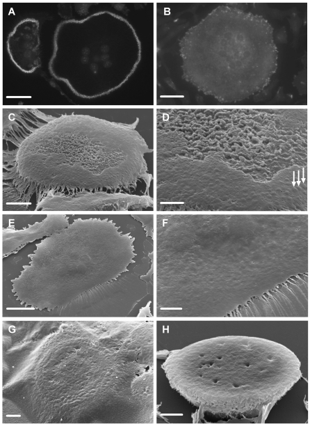
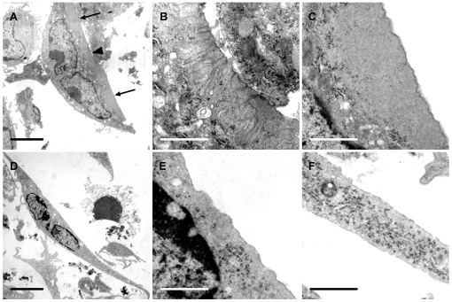
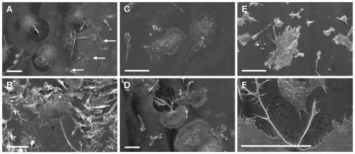
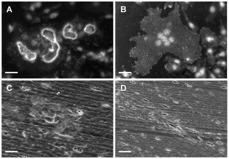
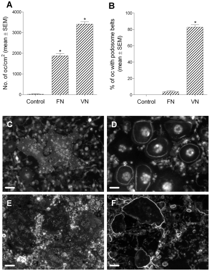
Methodology/principal findings: Vitronectin- but not fibronectin-coated coverslips induced murine osteoclasts to secrete tartrate-resistant acid phosphatase, as they do on bone. Osteoclasts incubated on vitronectin, unlike fibronectin, formed podosome belts on glass coverslips, and these were modulated by resorption-regulating cytokines. Podosome belts formed on vitronectin-coated surfaces whether the substrates were rough or smooth, rigid or flexible. We developed a novel approach whereby the substrate-apposed surface of cells can be visualized in the scanning electron microscope. With this approach, supported by transmission electron microscopy, we found that osteoclasts on vitronectin-coated surfaces show ruffled borders and clear zones characteristic of resorbing osteoclasts. Ruffles were obscured by a film if cells were incubated in the cathepsin inhibitor E64, suggesting that removal of the film represents substrate-degrading behavior. Analogously, osteoclasts formed resorption-like trails on vitronectin-coated substrates. Like bone resorption, these trails were dependent upon resorbogenic cytokines and were inhibited by E64. Bone mineral induced actin rings and surface excavation only if first coated with vitronectin. Fibronectin could not substitute in any of these activities, despite enabling adhesion and cell spreading.

Conclusions/significance: Our results show that ligands α(v)β(3) are not only necessary but sufficient for the induction of resorptive behavior in osteoclasts; and suggest that bone is recognized through its affinity for these ligands, rather than by its mechanical or topographical attributes, or through a putative 'mineral receptor'.


Conflict of interest statement

Competing Interests: The authors have declared that no competing interests exist.

Figures

Figure 1
Figure 1. Vitronectin induces TRAP release by osteoclasts.
Bone marrow-derived osteoclasts were sedimented in MEM/2.5% FCS onto coverslips that had been coated with the vitronectin or fibronectin at the concentrations shown. After 20 minutes, the coverslips were washed and incubated for 5 hours in MEM containing 2.5% FCS, M-CSF (50 ng/ml), RANKL (30 ng/ml) and IL-1α (10 ng/ml), and with/without salmon calcitonin (CT) (100 pg/ml). TRAP was then measured in the supernatant and lysate. n = 5 cultures per variable. A, B: *p<0.05 versus 0 group (A) or versus all other groups (B); ANOVA plus Dunnett's post-test. C: *p<0.05 versus no CT. ANOVA followed by Bonferroni post-test.
Figure 2
Figure 2. Podosome belt formation is regulated in a manner that parallels regulation of bone resorption.
Bone marrow-derived osteoclasts were sedimented in MEM/FCS onto glass coverslips. After 20minutes, the coverslips were washed and incubated for 5 hours in MEM/FCS in M-CSF (50 ng/ml) plus the agents shown (A), or MEM/FCS in M-CSF (50 ng/ml), RANKL (30 ng/ml) and IL-1α (10 ng/ml) with/without salmon calcitonin (B). A: OPG: 500 ng/ml; IL-1α: 10 ng/ml. *p<0.05 versus 0; +p<0.05 versus 0+IL-1µ, both ANOVA plus Dunnett's post-test; a: p<0.05 versus RANKL (30 ng/ml), ANOVA + Bonferroni post-test. n = 10 per variable. C-F: photomicrographs of phalloidin/DAPI preparations of cultures after incubation as above in: C: 0; D: RANKL (30 ng/ml); E: RANKL (30 ng/ml) + IL-1α; F: calcitonin (100 pg/ml). Scale bars  = 100 µm.
Figure 3
Figure 3. The effect of substrate roughness on the morphology of podosome belts.
Bone marrow-derived osteoclasts were sedimented in MEM/BSA onto Perspex that had been coated with vitronectin (50 µg/ml), or bone slices. Osteoclasts were incubated for 5 hours in MEM/BSA with M-CSF (50 ng/ml), RANKL (30 ng/ml) and IL-1α (10 ng/ml) on uncut Perspex surfaces (smooth), or on the surface of Perspex cut with the same saw as that used to prepare bone slices (rough). Phalloidin staining. Scale bars  = 50 µm.
Figure 4
Figure 4. Vitronectin induces podosome belt formation in osteoclasts.
Bone marrow-derived osteoclasts were sedimented in MEM/BSA onto glass coverslips that had been coated with the vitronectin or fibronectin at the concentrations shown. The coverslips were then washed and incubated for 5 hours in MEM/BSA with M-CSF (50 ng/ml), RANKL (30 ng/ml) and IL-1α (10 ng/ml) before fixation and staining for F-actin and DAPI. Vitronectin (A–C) induced a dose-dependent increase in adhesion and podosome belt formation in osteoclasts. In contrast, fibronectin (D–F), effectively induced adhesion of osteoclasts, but very few of the adherent cells developed podosome belts. *p<0.05 versus no adhesive ligand (ANOVA + Dunnett's). n = 20 fields (10x objective) per group. G, H: representative views of phalloidin/DAPI-stained osteoclasts incubated on vitronectin (G) and fibronectin (H). Osteoclasts on fibronectin are devoid of podosome belts. Scale bars  = 100 µm.
Figure 5
Figure 5. Vitronectin induces formation of ruffled border in osteoclasts.
Glass coverslips were coated with nail-varnish. This was then coated with vitronectin or fibronectin (50 µg/ml). Next, osteoclasts were incubated on this surface for 5 hours in MEM/BSA with M-CSF (50 ng/ml), RANKL (30 ng/ml) and IL-1α (10 ng/ml), with/without the cathepsin inhibitor E64 (3×10⁻⁷ M) or calcitonin (100 pg/ml). A,B: Phalloidin-stained preparations after incubation on vitronectin (A) or fibronectin (B). In A, individual podosomes can be discerned within the circumferential belt of podosomes. No podosomes are seen in the osteoclast that had been incubated on fibronectin. C–H: The discs of nail-varnish were separated from the glass coverslip, inverted onto a glass slide, dissolved in acetone, dehydrated in HMDS, and sputter-coated with gold for visualization in the SEM. C,D: undersurface of osteoclast incubated on vitronectin. The central area of the undersurface of the cell is filled with dense membrane folds, while the circumference lacks folds, but shows raised foci likely to represent podosomes (arrows). Note residual film of protein attached to the cell periphery. D: higher magnification of C. Note variation in density of membrane folds in the central area. E,F: Low and higher power view of undersurface of osteoclast incubated on fibronectin. The surface is relatively featureless, and lacks the domain organization apparent after incubation on vitronectin. G: The undersurface of osteoclasts was obscured by a protein film in cultures to which the cathepsin inhibitor E64 was added. Nevertheless, a peripheral belt of raised, podosome-like structures can be discerned through the film. H: The undersurface of osteoclasts incubated with calcitonin lacked membrane ruffles and podosome belts. Scale bars: A,B: 35 µm; C,F,H: 5 µm; D,G: 2 µm; E: 20 µm.
Figure 6
Figure 6. Vitronectin induces ruffled borders and clear zones in osteoclasts.
Osteoclasts were incubated for 5 hours in MEM/BSA with M-CSF (50 ng/ml), RANKL (30 ng/ml) and IL-1α (10 ng/ml) in 6-well plate wells coated with vitronectin or fibronectin (50 µg/ml), before raising into suspension with a cell scraper and preparation for TEM. A: Osteoclast incubated on vitronectin shows a central area of ruffled border (arrowhead) and a peripheral area free of organelles (‘clear zone’) (arrows). B, C: higher magnification of center (B) and lower portion (C) respectively of A, showing area of ruffled border (B) and clear zone (C); D–F: Osteoclast incubated on fibronectin shows well-spread appearance, but the undersurface lacked the membrane folds and clear zones seen in osteoclasts incubated on vitronectin. E and F are from central and lower portion of D respectively. Scale bars: A, D: 5 µm; B, C, E, F: 1 µm.
Figure 7
Figure 7. Vitronectin induces formation of resorption-like trails by osteoclasts on glass substrates.
Osteoclasts were incubated for 5 hours in MEM/BSA with M-CSF (50 ng/ml), RANKL (30 ng/ml) and IL-1α (10 ng/ml) (A, C–F), or M-CSF (B), together with the cathepsin inhibitor E64 (3×10⁻⁷ M) (C) or GM6001 (1.3×10⁻⁵ M) (D), on glass coverslips coated with vitronectin (A–D) or fibronectin (E, F) (50 µg/ml), before preparation for SEM. A: Osteoclasts incubated in resorption-inducing cytokines on vitronectin show well-defined cleared areas at the retreating margins of the cells. Macrophagic cells (some of which are identified by arrows, as the smaller cells, with leaf-like, rather than filopodial membrane folds) are not associated with cleared areas. B: Only occasional, and small, cleared areas were seen in cultures to which resorption-inducing cytokines had not been added. C: The formation of cleared areas was inhibited by the cysteine protease inhibitor E64. D: the MMP inhibitor GM6001 was without apparent effect. E, F: No cleared areas were seen at the retreating pole of osteoclasts incubated on fibronectin, although there was evidence of focal disturbance to the surface of the protein film in the region of filopodia. Scale bars  = 50 µm.
Figure 8
Figure 8. Vitronectin coating of anorganic bone enables podosome belt formation and resorption.
Osteoclasts were incubated for 5 hours in MEM/BSA with M-CSF (50 ng/ml), RANKL (30 ng/ml) and IL-1α (10 ng/ml) on slices of anorganic bone coated with vitronectin or fibronectin (50 µg/ml). Podosome belts and excavations were frequently seen on vitronectin-coated anorganic bone slices (A, C), but were never seen on fibronectin-coated anorganic bone slices (B, D). A, B: Phalloidin/DAPI staining; C, D: SEM images. Scale bars A–C: 25 µm; D: 50 µm.
Figure 9
Figure 9. Flexible substrates can induce podosome belt formation.
Osteoclasts were incubated for 5 hours in MEM/BSA with M-CSF (50 ng/ml), RANKL (30 ng/ml) and IL-1α (10 ng/ml) on PDMS sheets or silicone films coated with vitronectin or fibronectin (50 µg/ml). A,B: Osteoclasts adhered to fibronectin-coated PDMS sheets in large numbers, but podosome belts were very rare. In contrast, a similar proportion of osteoclasts formed podosome belts on vitronectin-coated PDMS sheets to our previous experience using rigid substrates. *p<0.05 versus control (ANOVA + Dunnett's); n = 10 per group. C–F: Representative views of osteoclasts on fibronectin-coated silicone film (C) or PDMS sheet (E)0; and vitronectin-coated silicone film (D) or PDMS sheet (F). Note that osteoclasts on fibronectin-coated substrates are well-spread but lack podosome belts. Phalloidin/DAPI staining. Scale bars: C,D: 50 µm; E,F: 100 µm.

Similar articles

Cited by

References

    1. Chambers TJ. Regulation of the differentiation and function of osteoclasts. J Pathol. 2000;192:4–13. - PubMed
    1. Suda T, Takahashi N, Udagawa N, Jimi E, Gillespie MT, et al. Modulation of osteoclast differentiation and function by the new members of the tumor necrosis factor receptor and ligand families. Endocr Rev. 1999;20:345–357. - PubMed
    1. Vaananen HK, Zhao H, Mulari M, Halleen JM. The cell biology of osteoclast function. J Cell Sci. 2000;113(Pt 3):377–381. - PubMed
    1. Stenbeck G. Formation and function of the ruffled border in osteoclasts. Semin Cell Dev Biol. 2002;13:285–292. - PubMed
    1. Fisher JE, Caulfield MP, Sato M, Quartuccio HA, Gould RJ, et al. Inhibition of Osteoclastic Bone-Resorption Invivo by Echistatin, an Arginyl-Glycyl-Aspartyl (Rgd)-Containing Protein. Endocrinology. 1993;132:1411–1413. - PubMed

Publication types