Genetic deletion of the desmosomal component desmoplakin promotes tumor microinvasion in a mouse model of pancreatic neuroendocrine carcinogenesis
- PMID: 20862307
- PMCID: PMC2940733
- DOI: 10.1371/journal.pgen.1001120
Genetic deletion of the desmosomal component desmoplakin promotes tumor microinvasion in a mouse model of pancreatic neuroendocrine carcinogenesis
Abstract
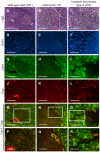
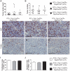
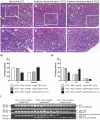
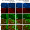
We used the RIP1-Tag2 (RT2) mouse model of islet cell carcinogenesis to profile the transcriptome of pancreatic neuroendocrine tumors (PNET) that were either non-invasive or highly invasive, seeking to identify pro- and anti-invasive molecules. Expression of multiple components of desmosomes, structures that help maintain cellular adhesion, was significantly reduced in invasive carcinomas. Genetic deletion of one of these desmosomal components, desmoplakin, resulted in increased local tumor invasion without affecting tumor growth parameters in RT2 PNETs. Expression of cadherin 1, a component of the adherens junction adhesion complex, was maintained in these tumors despite the genetic deletion of desmoplakin. Our results demonstrate that loss of desmoplakin expression and resultant disruption of desmosomal adhesion can promote increased local tumor invasion independent of adherens junction status.
Conflict of interest statement
The authors have declared that no competing interests exist.
Figures


Similar articles
-
Genetic ablation of Bcl-x attenuates invasiveness without affecting apoptosis or tumor growth in a mouse model of pancreatic neuroendocrine cancer.PLoS One. 2009;4(2):e4455. doi: 10.1371/journal.pone.0004455. Epub 2009 Feb 11. PLoS One. 2009. PMID: 19209227 Free PMC article.
-
Altered desmoplakin expression at transcriptional and protein levels provides prognostic information in human oropharyngeal cancer.Hum Pathol. 2009 Sep;40(9):1320-9. doi: 10.1016/j.humpath.2009.02.002. Epub 2009 Apr 22. Hum Pathol. 2009. PMID: 19386346
-
Targeting of p0071 to desmosomes and adherens junctions is mediated by different protein domains.J Cell Sci. 2003 Apr 1;116(Pt 7):1219-33. doi: 10.1242/jcs.00275. J Cell Sci. 2003. PMID: 12615965
-
In vivo function of desmosomes.J Dermatol. 2004 Mar;31(3):171-87. doi: 10.1111/j.1346-8138.2004.tb00654.x. J Dermatol. 2004. PMID: 15187337 Review.
-
Animal models and cell lines of pancreatic neuroendocrine tumors.Pancreas. 2013 Aug;42(6):912-23. doi: 10.1097/MPA.0b013e31827ae993. Pancreas. 2013. PMID: 23851429 Review.
Cited by
-
Desmosomes: regulators of cellular signaling and adhesion in epidermal health and disease.Cold Spring Harb Perspect Med. 2014 Nov 3;4(11):a015297. doi: 10.1101/cshperspect.a015297. Cold Spring Harb Perspect Med. 2014. PMID: 25368015 Free PMC article. Review.
-
Structure, function, and regulation of desmosomes.Prog Mol Biol Transl Sci. 2013;116:95-118. doi: 10.1016/B978-0-12-394311-8.00005-4. Prog Mol Biol Transl Sci. 2013. PMID: 23481192 Free PMC article. Review.
-
The attributes of plakins in cancer and disease: perspectives on ovarian cancer progression, chemoresistance and recurrence.Cell Commun Signal. 2021 May 17;19(1):55. doi: 10.1186/s12964-021-00726-x. Cell Commun Signal. 2021. PMID: 34001250 Free PMC article. Review.
-
Death-associated protein kinase 3 modulates migration and invasion of triple-negative breast cancer cells.PNAS Nexus. 2024 Sep 12;3(9):pgae401. doi: 10.1093/pnasnexus/pgae401. eCollection 2024 Sep. PNAS Nexus. 2024. PMID: 39319326 Free PMC article.
-
Desmosomes in acquired disease.Cell Tissue Res. 2015 Jun;360(3):439-56. doi: 10.1007/s00441-015-2155-2. Epub 2015 Mar 21. Cell Tissue Res. 2015. PMID: 25795143 Free PMC article. Review.
References
-
- Hanahan D, Weinberg RA. The hallmarks of cancer. Cell. 2000;100:57–70. - PubMed
-
- Christofori G. New signals from the invasive front. Nature. 2006;441:444–450. - PubMed
-
- Shinohara M, Hiraki A, Ikebe T, Nakamura S, Kurahara S, et al. Immunohistochemical study of desmosomes in oral squamous cell carcinoma: correlation with cytokeratin and E-cadherin staining, and with tumour behaviour. J Pathol. 1998;184:369–381. - PubMed
Publication types
MeSH terms
Substances
Grants and funding
LinkOut - more resources
Full Text Sources
Medical
Molecular Biology Databases
Miscellaneous

