. 2010 Sep 16;6(9):e1001108.
doi: 10.1371/journal.ppat.1001108.

Generation of genic diversity among Streptococcus pneumoniae strains via horizontal gene transfer during a chronic polyclonal pediatric infection

Affiliations

Generation of genic diversity among Streptococcus pneumoniae strains via horizontal gene transfer during a chronic polyclonal pediatric infection

N Luisa Hiller et al. PLoS Pathog. .

Abstract

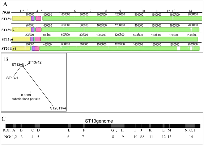
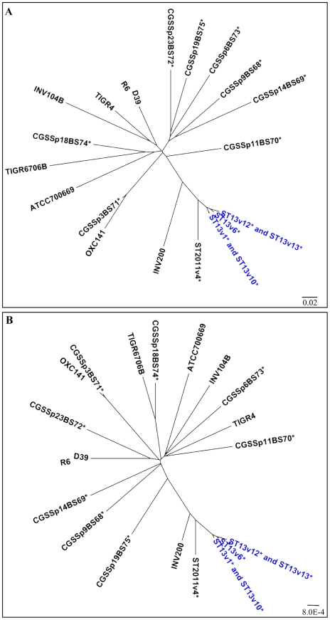
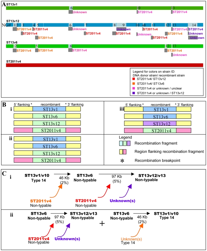
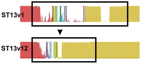
Although there is tremendous interest in understanding the evolutionary roles of horizontal gene transfer (HGT) processes that occur during chronic polyclonal infections, to date there have been few studies that directly address this topic. We have characterized multiple HGT events that most likely occurred during polyclonal infection among nasopharyngeal strains of Streptococcus pneumoniae recovered from a child suffering from chronic upper respiratory and middle-ear infections. Whole genome sequencing and comparative genomics were performed on six isolates collected during symptomatic episodes over a period of seven months. From these comparisons we determined that five of the isolates were genetically highly similar and likely represented a dominant lineage. We analyzed all genic and allelic differences among all six isolates and found that all differences tended to occur within contiguous genomic blocks, suggestive of strain evolution by homologous recombination. From these analyses we identified three strains (two of which were recovered on two different occasions) that appear to have been derived sequentially, one from the next, each by multiple recombination events. We also identified a fourth strain that contains many of the genomic segments that differentiate the three highly related strains from one another, and have hypothesized that this fourth strain may have served as a donor multiple times in the evolution of the dominant strain line. The variations among the parent, daughter, and grand-daughter recombinant strains collectively cover greater than seven percent of the genome and are grouped into 23 chromosomal clusters. While capturing in vivo HGT, these data support the distributed genome hypothesis and suggest that a single competence event in pneumococci can result in the replacement of DNA at multiple non-adjacent loci.


Conflict of interest statement

The authors have declared that no competing interests exist.

Figures

Figure 1
Figure 1. Demonstration of recombination events among Streptococcus pneumoniae genomes isolated from a single patient.
(A) MAUVE alignment of the WGSs of four S. pneumoniae isolates showing four distinct strains. Colored blocks highlight regions that are homologous and free from genomic rearrangement. White areas illustrate degrees of average conservation that exist within corresponding genome regions (more white  =  less conservation). Neighbor group (NG) numbers above the schematic denote the positions of clustered SNP groupings. (B) Maximum likelihood phylogenetic tree (given the best fit model  =  F84+G4) expressing the relationship between the four genomes and demonstrating that ST2011v4 is the most divergent of these strains and that differences exist among the ST13 strains. (C) Schematic showing the relative position of recombination events (gray boxes) within the ST13 genome (black), as predicted by RDP3 and NG analyses.
Figure 2
Figure 2. High degree of genomic similarity among ST13 strains.
Grouping of 22 S. pneumoniae isolates based on (A) numbers of distributed genes and (B) numbers of variable core alleles. These graphs provide a measure of the genic or alleic distance among isolates without making any inference regarding their phylogeny, since the high rates of recombination within the population can interfere with population-wide phylogenetic results. The high degree of similarity among the ST13 strains isolated from patient 19 suggests that these strains have evolved within this patient and are not the result of independent infections. Further details on the strains are described in Hiller et al .
Figure 3
Figure 3. Identification of DNA donors for recombinant ST13 isolates.
(A) RDP3-generated schematic of four genomes indicating evidence of recombination with neighbor group (NG) results superimposed. Light gray boxes within the whole genome sequence (WGS) schematic represent recombination events, and corresponding NG numbers have been inserted. Dark gray boxes are positioned below likely recombinant strains and are labeled with the most likely DNA donor for each transferred region. (B) Schematic illustrating the various patterns found in both the recombinant regions and surrounding areas. (i) Recombination fragments that are identical among ST2011v4, ST13v6, and ST13v12 while the surrounding regions are identical only among the ST13 strains (but contain SNPs relative to ST2011v4) (representative of NGs 5 and12 and corresponding to RDP D and L); (ii) Recombination fragments that are identical only between ST2011v4 and ST13v12 while the surrounding regions are identical only among the ST13 strains (representative of NGs 1,3,4,7, and 11 and corresponding to RDP part of A, B,C, F and I); (iii) Recombination fragments that differ between ST13v12 and all other strains (representative of NGs 9 and 14 and corresponding to RDP part of G and N, O and P). (C) Schematic outlining possible recombination events that may have led to the creation of the strains isolated from the patient. (i) most and (ii) next-most parsimonious. Black arrows move from the major parental strain to the recombinant, colors highlight likely DNA donor strains. The conservative estimate for the size of the recombination fragments is marked below the arrows with the corresponding percentage of the WGS in parenthesis.
Figure 4
Figure 4. Illustration of a region transferred by HGT.
MAUVE alignment of the genome region of ST13v1 and ST13v12 corresponding to NG14 (containing RDP N, O and P). Colored blocks represent NG14 regions that are homologous; a similarity plot inside each block portrays the average degree of conservation in that region. Black boxes bound sites displaying the most obvious differences between the 57 Kb NG14 region of ST13v1 and the 34 Kb NG14 region of ST13v12.
Figure 5
Figure 5. Phenotypic differences among ST13 strains.
Biofilm development after 5 days on polystyrene plates by clinical isolates (i) ST13v1, (ii) ST13v6, and (iii) ST13v12. Images are maximum projections of reconstructed confocal stacks consisting of a series of x–y sections. Cells were stained with Syto59 . Scale bar  = 30 µm.

Similar articles

Cited by

References

    1. Gogarten JP, Townsend JP. Horizontal gene transfer, genome innovation and evolution. Nat Rev Microbiol. 2005;3:679–687. - PubMed
    1. Tettelin H, Masignani V, Cieslewicz MJ, Donati C, Medini D, et al. Genome analysis of multiple pathogenic isolates of Streptococcus agalactiae: implications for the microbial “pan-genome”. Proc Natl Acad Sci U S A. 2005;102:13950–13955. - PMC - PubMed
    1. Hogg JS, Hu FZ, Janto B, Boissy R, Hayes J, et al. Characterization and modeling of the Haemophilus influenzae core and supragenomes based on the complete genomic sequences of Rd and 12 clinical nontypeable strains. Genome Biol. 2007;8:R103. - PMC - PubMed
    1. Hiller NL, Janto B, Hogg JS, Boissy R, Yu S, et al. Comparative Genomic Analyses of Seventeen Streptococcus pneumoniae Strains: Insights into the Pneumococcal Supragenome. J Bacteriol. 2007;189:8186–8195. - PMC - PubMed
    1. Smith JM, Dowson CG, Spratt BG. Localized sex in bacteria. Nature. 1991;349:29–31. - PubMed

Publication types

MeSH terms