. 2010 Sep 16;6(9):e1001106.
doi: 10.1371/journal.ppat.1001106.

The HA and NS genes of human H5N1 influenza A virus contribute to high virulence in ferrets

Affiliations

The HA and NS genes of human H5N1 influenza A virus contribute to high virulence in ferrets

Hirotaka Imai et al. PLoS Pathog. .

Abstract

Highly pathogenic H5N1 influenza A viruses have spread across Asia, Europe, and Africa. More than 500 cases of H5N1 virus infection in humans, with a high lethality rate, have been reported. To understand the molecular basis for the high virulence of H5N1 viruses in mammals, we tested the virulence in ferrets of several H5N1 viruses isolated from humans and found A/Vietnam/UT3062/04 (UT3062) to be the most virulent and A/Vietnam/UT3028/03 (UT3028) to be avirulent in this animal model. We then generated a series of reassortant viruses between the two viruses and assessed their virulence in ferrets. All of the viruses that possessed both the UT3062 hemagglutinin (HA) and nonstructural protein (NS) genes were highly virulent. By contrast, all those possessing the UT3028 HA or NS genes were attenuated in ferrets. These results demonstrate that the HA and NS genes are responsible for the difference in virulence in ferrets between the two viruses. Amino acid differences were identified at position 134 of HA, at positions 200 and 205 of NS1, and at positions 47 and 51 of NS2. We found that the residue at position 134 of HA alters the receptor-binding property of the virus, as measured by viral elution from erythrocytes. Further, both of the residues at positions 200 and 205 of NS1 contributed to enhanced type I interferon (IFN) antagonistic activity. These findings further our understanding of the determinants of pathogenicity of H5N1 viruses in mammals.


Conflict of interest statement

The authors have declared that no competing interests exist.

Figures
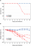
Figure 1
Figure 1. UT3062 differs from UT3028 in its virulence in ferrets.
(A) Survival of ferrets infected with 107 PFU of UT3062 (n = 6) and UT3028 (n = 3) is indicated. (B) Percent body weight change of each virus-infected ferret is shown.
Figure 2
Figure 2. The HA and NS genes contribute to virulence in ferrets.
Ferrets were infected with 107 PFU of reassortant viruses that combine the genes of UT3062 (red) and UT3028 (white), generated by reverse genetics, and with the parental UT3062 and UT3028 viruses. Lethality, mean maximum weight change ± standard deviations (SD), and the number of ferrets used (n) are indicated. Animals with greater than 20% weight loss, hemorrhage from any body orifice, or inability to remain upright were euthanized. Pol, polymerase complex. HA+NA, surface glycoproteins, HA and NA.
Figure 3
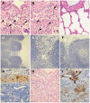
Figure 3. Pathological findings in ferrets infected with UT3062 or 3062(HA+NS).
(A) Lungs of ferret infected with UT3062. Infiltration of eosinophils (arrowheads and insert) was obvious around the bronchi 1 day p.i. (B) Lungs of ferrets infected with UT3028. Neutrophils (arrowheads and insert) were apparent in the lung lesion 1 day p.i. (C) The bronchial area of normal appearance has clear air spaces. (D) Immunostaining of tracheobronchial lymph node in a ferret infected with UT3062. Virus antigen can be seen in the sinusoid area (brown pigment) 1 day p.i. (E) Higher magnification of Figure 3D's broken-line square area. Reticular cell-like cells in the sinusoid stain positive for virus antigen (brown pigment). (F) The lymph node with normal appearance did not contain brown pigments. (G) Immunostaining of tracheobronchial lymph node in a ferret infected with 3062(HA+NS). Many viral antigen-positive cells are visible in the sinusoid (brown pigment) 7 days p.i. (H) Lung lesion in a ferret infected with 3062(HA+NS). Prominent inflammatory infiltrates and hemorrhage are detected 7 days p.i. by H&E staining. (I) Immunostaining of Figure 3H lesion. Heavy staining of virus antigen can be seen in the bronchial epithelium and alveolar cells (brown pigments) 7 days p.i.
Figure 4
Figure 4. Distribution of lung lesions and viral antigens in ferrets.
(A) Lungs of ferrets infected with UT3028. 1, 3, 5, and 7 days p.i. (B) Lungs of ferrets infected with UT3062. 1, 3, 5, and 7 days p.i. (C) Lungs of ferrets infected with 3062(HA+NS). 3 and 7 days p.i. Viral antigens in lung lesions, +++; many, ++; moderate, +; few, ±; scant, −; not detected. Distributions of lung lesions and viral antigens were confirmed by microscopic examination.
Figure 5
Figure 5. UT3062 and UT3028 differ in their virus elution ability from erythrocyte.
Two-fold dilutions of virus containing HA titers of 1∶64 were incubated with equal volumes of 0.55% chicken erythrocytes at 4°C for 1 h. They were then incubated at 37°C and the reduction in HA titers was recorded for 20 h.
Figure 6
Figure 6. The 3062 NS gene promotes IFN antagonism.
Mv1Lu cells were infected with each virus at an MOI of 1.25. Supernatants from virus-infected cells were harvested 12–24 h p.i. After inactivation of viral infectivity, the supernatants were added to fresh Mv1Lu cells and incubated for 22 h. The pretreated Mv1Lu cells were then infected with VSV and infectivity was assessed by plaque assays. N200S represents the virus expressing mutant NS1 protein in which asparagine was substituted to serine and mutant NS2 protein in which threonine was substituted to alanine on the UT3028 backbone. G205R represents the virus expressing mutant NS1 protein in which glycine was substituted to arginine and mutant NS2 in which methionine was substituted to isoleucine on the UT3028 backbone. As a control, we used a recombinant influenza virus expressing an RNA-binding- and IFN antagonism-defective NS1 protein within which two basic amino acids were substituted to alanines (R38A/K41A) on the UT3062 backbone. The data are shown as mean VSV titers (log10 PFU/ml) ± SD of triplicate values obtained in a single experiment, and are representative of two independent experiments.

References

    1. Claas E, Osterhaus A, van Beek R, De Jong J, Rimmelzwaan G, et al. Human influenza A H5N1 virus related to a highly pathogenic avian influenza virus. Lancet. 1998;351:472–477. - PubMed
    1. Subbarao K, Klimov A, Katz J, Regnery H, Lim W, et al. Characterization of an avian influenza A (H5N1) virus isolated from a child with a fatal respiratory illness. Science. 1998;279:393–396. - PubMed
    1. Barnard D. Animal models for the study of influenza pathogenesis and therapy. Antiviral Res. 2009;82:A110–122. - PMC - PubMed
    1. Hatta M, Gao P, Halfmann P, Kawaoka Y. Molecular basis for high virulence of Hong Kong H5N1 influenza A viruses. Science. 2001;293:1840–1842. - PubMed
    1. Govorkova E, Rehg J, Krauss S, Yen H, Guan Y, et al. Lethality to ferrets of H5N1 influenza viruses isolated from humans and poultry in 2004. J Virol. 2005;79:2191–2198. - PMC - PubMed

Publication types

MeSH terms

Substances