

Identification of myocardial damage in systemic sclerosis: a nuclear cardiology approach
- PMID: 20862337
- PMCID: PMC2939403
- DOI: 10.1155/2010/496509
Identification of myocardial damage in systemic sclerosis: a nuclear cardiology approach
Abstract
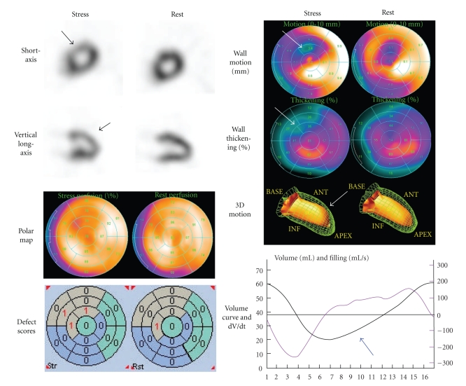
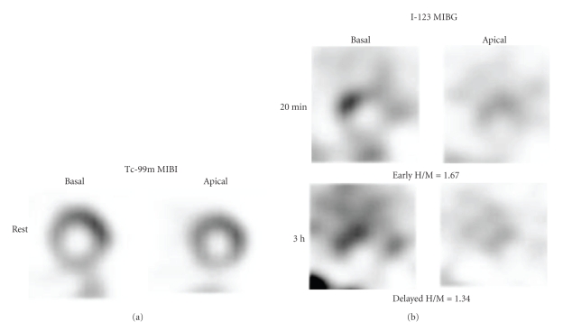
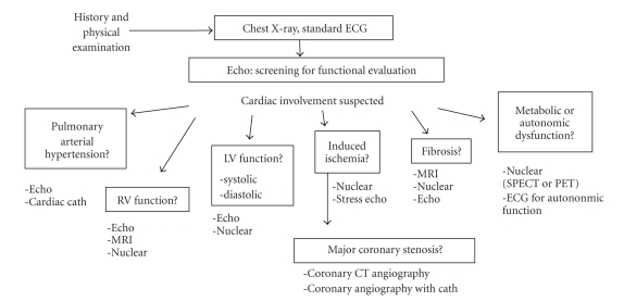
Myocardial involvement is an important prognostic factor in patients with systemic sclerosis, and early diagnosis and staging of the disease have been sought after. Since myocardial damage is characterized by connective tissue disease, including fibrosis and diffuse vascular lesions or microcirculation, nuclear myocardial perfusion imaging has been a promising option for evaluating myocardial damages in early stages. In addition to the conventional stress-rest perfusion imaging, the current use of quantitative electrocardiographic gated imaging has contributed to more precise evaluation of cardiac perfusion, ventricular wall motion, and diastolic function, all of which have enhanced diagnostic ability of evaluating myocardial dysfunction. Abnormal sympathetic imaging with Iodine-123 metaiodobenzylguanidine might be another option for identifying myocardial damage. This paper deals with approaches from nuclear cardiology to detect perfusion and functional abnormality as an early sign of myocardial involvement as well as possible prognostic values in patients with abnormal imaging results. The role of nuclear cardiology in the era of multiple imaging modalities is discussed.
Figures
References
-
- D’Angelo WA, Fries JF, Masi. AT, Shulman LE. Pathologic observations in systemic sclerosis (scleroderma). A study of fifty-eight autopsy cases and fifty-eight matched controls. The American Journal of Medicine. 1969;46(3):428–440. - PubMed
-
- Medsger TA, Jr., Masi AT, Rodnan GP, Benedek TG, Robinson H. Survival with systemic sclerosis (scleroderma). A life-table analysis of clinical and demographic factors in 309 patients. Annals of Internal Medicine. 1971;75(3):369–376. - PubMed
-
- LeRoy EC, Black C, Fleischmajer R, et al. Scleroderma (systemic sclerosis): classification, subsets and pathogenesis. Journal of Rheumatology. 1988;15(2):202–205. - PubMed
-
- Clements PJ, Lachenbruch PA, Seibold JR, et al. Skin thickness score in systemic sclerosis: an assessment of interobserver variability in 3 independent studies. Journal of Rheumatology. 1993;20(11):1892–1896. - PubMed
-
- Furst DE, Clements PJ, Steen VD, et al. The modified rodnan skin score is an accurate reflection of skin biopsy thickness in systemic sclerosis. Journal of Rheumatology. 1998;25(1):84–88. - PubMed
LinkOut - more resources
Full Text Sources
