. 2010 Dec;67(12):755-72.
doi: 10.1002/cm.20485. Epub 2010 Nov 2.

Assembly of filopodia by the formin FRL2 (FMNL3)

Affiliations

Assembly of filopodia by the formin FRL2 (FMNL3)

Elizabeth S Harris et al. Cytoskeleton (Hoboken). 2010 Dec.

Abstract

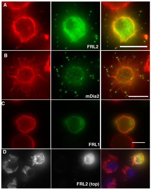
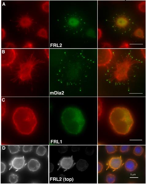
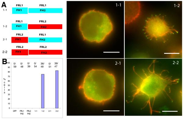
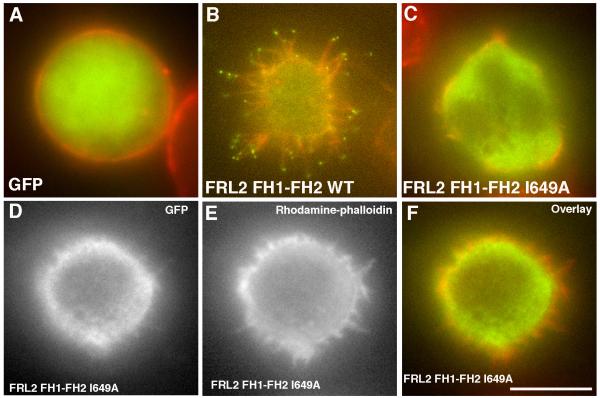
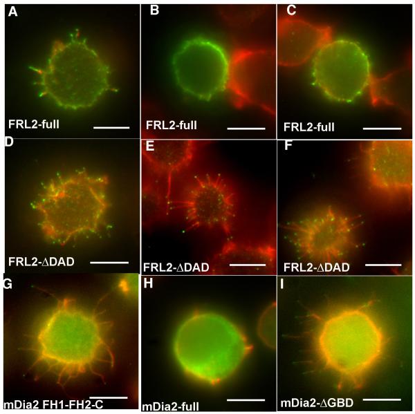
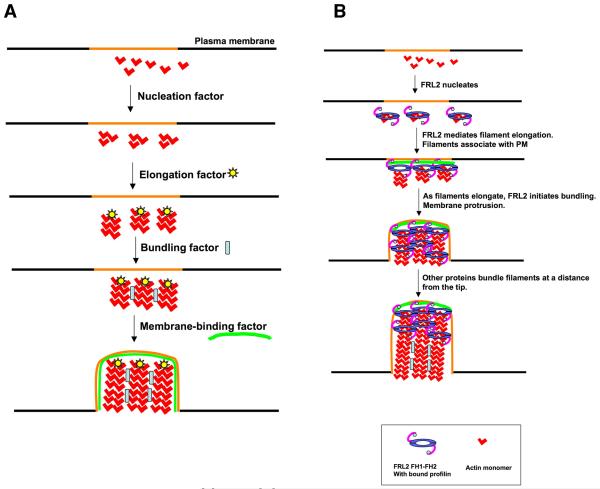
Actin-dependent finger-like protrusions such as filopodia and microvilli are widespread in eukaryotes, but their assembly mechanisms are poorly understood. Filopodia assembly requires at least three biochemical activities on actin: actin filament nucleation, prolonged actin filament elongation, and actin filament bundling. These activities are shared by several mammalian formin proteins, including mDia2, FRL1 (also called FMNL1), and FRL2 (FMNL3). In this paper, we compare the abilities of constructs from these three formins to induce filopodia. FH1-FH2 constructs of both FRL2 and mDia2 stimulate potent filopodia assembly in multiple cell types, and enrich strongly at filopodia tips. In contrast, FRL1 FH1-FH2 lacks this activity, despite possessing similar biochemical activities and being highly homologous to FRL2. Chimeric FH1-FH2 experiments between FRL1 and FRL2 show that, while both an FH1 and an FH2 are needed, either FH1 domain supports filopodia assembly but only FRL2's FH2 domain allows this activity. A mutation that compromises FRL2's barbed end binding ability abolishes filopodia assembly. FRL2's ability to stimulate filopodia assembly is not altered by additional domains (GBD, DID, DAD), but is significantly reduced in the full-length construct, suggesting that FRL2 is subject to inhibitory regulation. The data suggest that the FH2 domain of FRL2 possesses properties not shared by FRL1 that allow it to generate filopodia.


Figures

Figure 1
Figure 1. FRL2 and mDia2 FH1-FH2 domain-containing constructs induce filopodia in Jurkat cells
GFP-fusion constructs containing the FH1-FH2 domains of FRL2 (A, FH1-FH2 construct), mDia2 (B, FH1-FH2-C construct) or FRL1 (C, FH1-FH2-C construct) were expressed in Jurkat cells for six hours, followed by formaldehyde fixation and staining with rhodamine-phalloidin and DAPI. Panels A-C represent cells that were allowed to adhere to poly-L-lysine coated coverslips prior to fixation. Panel D represents cells that were transfected with FRL2 FH1-FH2-C construct, then fixed in suspension prior to adhesion to poly-L-lysine coated coverslips. Scale bar represents 5 μm.
Figure 2
Figure 2. FRL2 and mDia2 FH1-FH2 domain-containing constructs induce filopodia in 300.19 cells
GFP-fusion constructs containing the FH1-FH2 domains of FRL2 (A, FH1-FH2), mDia2 (B, FH1-FH2-C) or FRL1 (C, FH1-FH2-C) were expressed in 300.19 cells for six hours, followed by formaldehyde fixation and staining with rhodamine-phalloidin and DAPI. Panels A-C represent cells that were allowed to adhere to poly-L-lysine coated coverslips prior to fixation. Panel D represents cells that were transfected with FRL2 FH1-FH2-C then fixed in suspension prior to adhesion to poly-L-lysine coated coverslips. Scale bars represent 5 μm.
Figure 3
Figure 3. Dynamics of FRL2 FH1-FH2 induced filopodial protrusion in HeLa cells
HeLa cells were transfected with GFP-FRL2 FH1-FH2 (green) and mRFP-utropin-CH (red, a gift from Bill Bement) to label actin filaments. Micrographs were acquired at 10 sec intervals as Z-series and maximum intensity projections generated. The montage shows selected frames from Movie 3. Two filopodia actively protrude in this sequence. The white arrows mark the initial positions of the GFP-enriched filopodial tips, and remain in the same positions in subsequent frames to show progressive elongation. Times are given in sec.
Figure 4
Figure 4. The FH2 domain of FRL2 is essential for filopodia assembly
A) Chimeric constructs used in this experiment, expressed with an N-terminal GFP tag. B) Quantification of filopodia phenotype in Jurkat cells, compiled from three independent experiments. Numbers at the top of each column represent number of positive cells for microvilli/total number of cells counted. C-F) Examples of individual Jurkat cells for each of the chimeras. Scale bars, 5 μm.
Figure 5
Figure 5. I649A mutation disrupts barbed end binding but not bundling by FRL2
A) Sedimentation velocity analytical ultracentrifugation of 24 μM wild-type (black) or I649A mutant (red) FRL2 FH1-FH2. One major species sediments at 4.25 S. B) Polymerization of 2 μM actin monomers (10% pyrene-labeled) in the presence of the indicated concentrations of mDia1 or FRL2 FH1-FH2. C) Elongation rates of 1 μM actin monomers (10% pyrene-labeled) from phalloidin-stabilized actin filaments (1.5 μM) in the presence of the indicated concentrationsof wild-type or I649A FRL2 FH1-FH2. Inset shows raw elongation curves in the presence of 30 nM of the indicated construct. D) Low-speed pelleting assays of actin filament bundling by FRL2 FH1-FH2. Pre-polymerized actin filaments (2 μM) were mixed with the indicated nM concentrations of FRL2 FH1-FH2 construct, then centrifuged at 16,000xg for 10 min. Pellet fractions (representing bundled filaments with bound FRL2) are shown.
Figure 6
Figure 6. The FRL2 barbed end binding mutant is defective in filopodia assembly
Jurkat cells were transfected withGFP (A), GFP-FRL2 FH1-FH2 (B), or GFP-FRL2 FH1-FH2 I649A (C-F). After 6 hrs, the cells were plated onto poly-lysine-coated coverslips for 10 min, fixed, and stained with rhodamine-phalloidin. GFP, green. Rhodamine-phalloidin, red. Panels D, E and F show an I649A-transfected cell that displays short actin-rich protrusions. Panel D (GFP) shows that there is no enrichment of I649A to the tips of these structures, In contrast to the intense enrichment for wild-type (panel B). The cells displaying the phenotype shown in D-F make up less than 20% of the population. Scale bar, 5 μm.
Figure 7
Figure 7. Effects of additional domains on FRL2-induced filopodia
Jurkat cells were electroporated with GFP-fusion constructs of varying lengths of FRL2, then fixed and stained with rhodamine-phalloidin. A) Bar diagram of FRL2, showing the lengths of each construct. The FH1-FH2 construct is 504-945, FH1-FH2-C is 504-1027, DeltaN45 is 46-1027, Full is 1-1027, and ΔDAD.is 1-987. Domain boundaries are: GBD - 45-236; DID - 79-395; DD (dimerization domain) 398-452; FH1 - 505-548; FH2 - 561-945; DAD - 991-1000. The basis for the boundary predictions is given in Materials and Methods. B) - D) are micrographs of individual cells transfected with each construct. See Table 1 for quantification of phenotype. E) Filopodial lengths for the three constructs tested. Values for minimum and maximumlengths (in μm) and number of filopodia measured are as follows. FH1-FH2: 0.3 μm, 7.5 μm, and n= 109. FH1-FH2-C: 0.6, 9.4, and 70. DeltaN45: 0.6, 11.6, and 115. Scale bars represent 5 μm.
Figure 8
Figure 8. Full-length FRL2 has reduced ability to assemble filopodia
A-F) Jurkat cells were transfected with un-tagged constructs containing either full-length FRL2 (A-C), or FRL2 DDAD (D-F). After 6 hrs, cells were plated onto poly-lysine for 10 min, fixed, and stained with rhodamine-phalloidin (red) and anti-FRL2 followed by fluorescently labeled secondary antibody (green). Three examples of transfected cells are shown for each FRL2 construct. A) shows an example of a full-length FRL2 cell scored “positive” for filopodia, whereas B) and C) are “negative”. D)-F) are all examples of “positive” cells transfected with FRL2 ΔDAD. G-H) Jurkat cells transfected with GFP-fusions of mDia2 constructs. G) is mDia2 FH1-FH2-C, H) is mDia2 full-length, I) is mDia2-ΔGBD. Scale bars represent 5 μm.
Figure 9
Figure 9. Filopodia assembly models
A) Schematic representations of four biochemical activities needed for filopodia assembly from a designated region of the plasma membrane (orange). Theoretically, membrane binding could occur at any step, though we include it as the last step. Also, membrane binding could occur at the tips and/or the sides of filopodia. B) Working model for FRL2 FH1-FH2-mediated filopodia assembly, taking into account its known biochemical activities of nucleation acceleration, elongation regulation, and bundling. FRL2 FH1-FH2 nucleates filaments. Barbed end-bound FRL2 FH1-FH2 facilitates filament elongation through profilin. Additional FRL2 molecules mediate bundling near the tip. As the filaments elongate, other proteins bundle the filaments further from the tip. An unknown factor mediates membrane association. We depict membrane association at the elongation step, but it could occur during another step.

References

    1. Berg JS, Derfler BH, Pennisi CM, Corey DP, Cheney RE. Myosin-X, a novel myosin with pleckstrin homology domains, associates with regions of dynamic actin. J Cell Sci. 2000;113(Pt 19):3439–3451. - PubMed
    1. Block J, Stradal TE, Hanisch J, Geffers R, Kostler SA, Urban E, Small JV, Rottner K, Faix J. Filopodia formation induced by active mDia2/Drf3. J Microsc. 2008;231(3):506–17. - PubMed
    1. Bohil AB, Robertson BW, Cheney RE. Myosin-X is a molecular motor that functions in filopodia formation. Proc Natl Acad Sci U S A. 2006;103(33):12411–12416. - PMC - PubMed
    1. Brandt DT, Marion S, Griffiths G, Watanabe T, Kaibuchi K, Grosse R. Dia1 and IQGAP1 interact in cell migration and phagocytic cup formation. J Cell Biol. 2007;178(2):193–200. - PMC - PubMed
    1. Burkel BM, von Dassow G, Bement WM. Versatile fluorescent probes for actin filaments based on the actin-binding domain of utrophin. Cell Motil Cytoskeleton. 2007;64(11):822–832. - PMC - PubMed

Publication types

LinkOut - more resources