. 2010 Oct 26;4(10):5887-96.
doi: 10.1021/nn102055s.

A reexamination of active and passive tumor targeting by using rod-shaped gold nanocrystals and covalently conjugated peptide ligands

Affiliations

A reexamination of active and passive tumor targeting by using rod-shaped gold nanocrystals and covalently conjugated peptide ligands

Xiaohua Huang et al. ACS Nano. .

Erratum in

  • ACS Nano. 2011 Aug 23;5(8):6765

Abstract

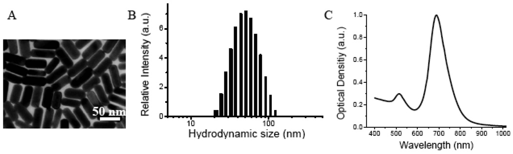
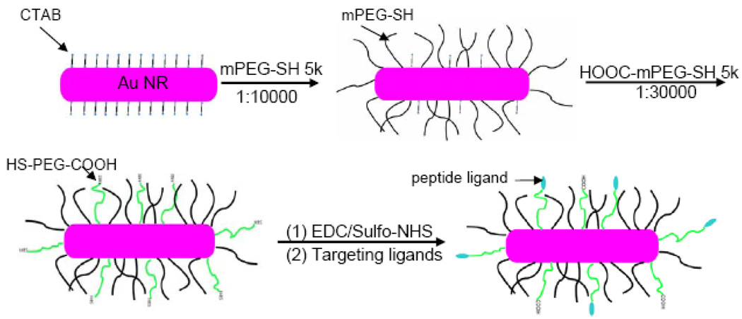
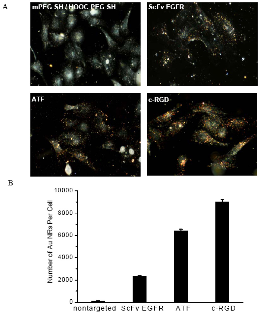
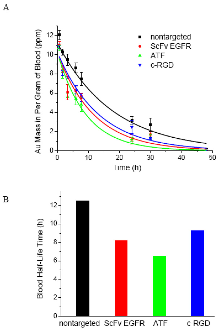
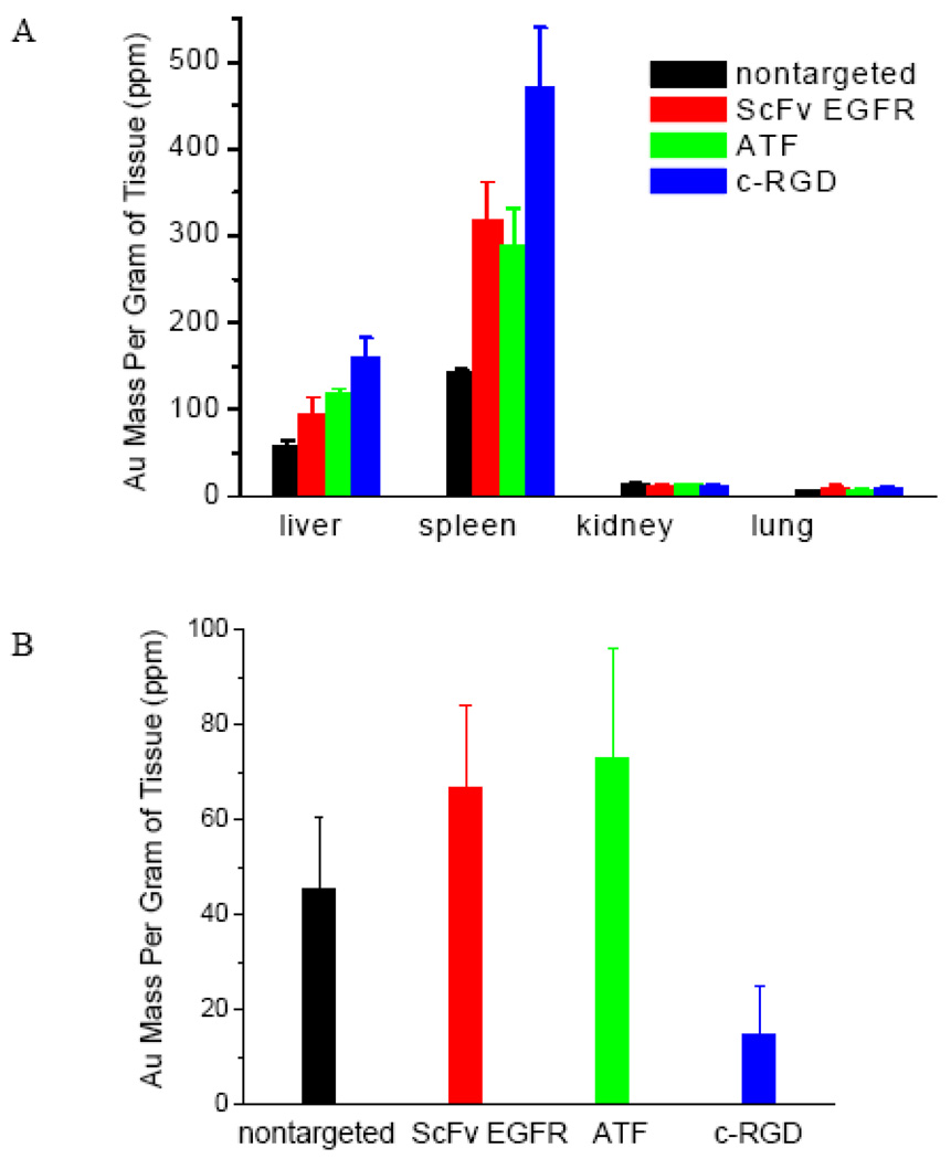
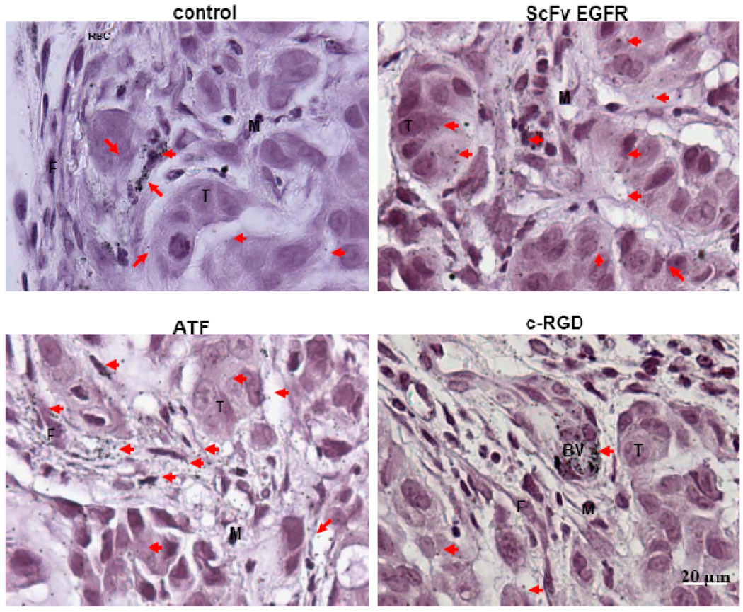
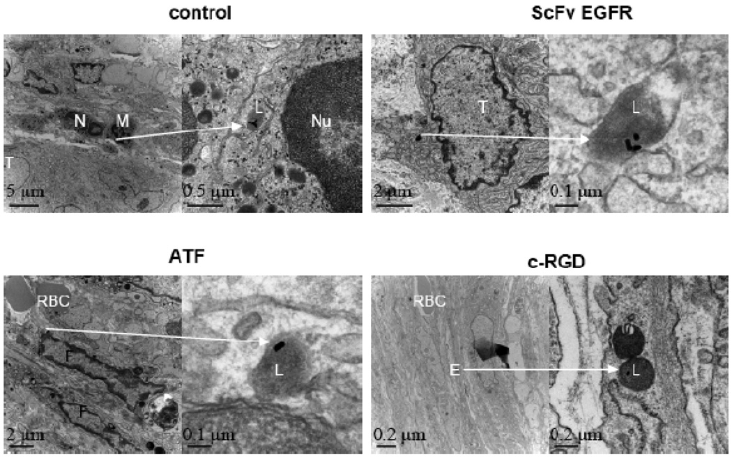
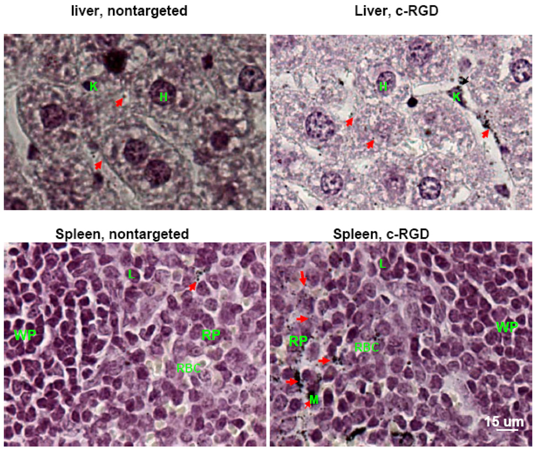
The targeted delivery of nanoparticles to solid tumors is one of the most important and challenging problems in cancer nanomedicine, but the detailed delivery mechanisms and design principles are still not well understood. Here we report quantitative tumor uptake studies for a class of elongated gold nanocrystals (called nanorods) that are covalently conjugated to tumor-targeting peptides. A major advantage in using gold as a "tracer" is that the accumulated gold in tumors and other organs can be quantitatively determined by elemental mass spectrometry (gold is not a natural element found in animals). Thus, colloidal gold nanorods are stabilized with a layer of polyethylene glycols (PEGs) and are conjugated to three different ligands: (i) a single-chain variable fragment (ScFv) peptide that recognizes the epidermal growth factor receptor (EGFR); (ii) an amino terminal fragment (ATF) peptide that recognizes the urokinase plasminogen activator receptor (uPAR); and (iii) a cyclic RGD peptide that recognizes the a(v)β(3) integrin receptor. Quantitative pharmacokinetic and biodistribution data show that these targeting ligands only marginally improve the total gold accumulation in xenograft tumor models in comparison with nontargeted controls, but their use could greatly alter the intracellular and extracellular nanoparticle distributions. When the gold nanorods are administered via intravenous injection, we also find that active molecular targeting of the tumor microenvironments (e.g., fibroblasts, macrophages, and vasculatures) does not significantly influence the tumor nanoparticle uptake. These results suggest that for photothermal cancer therapy, the preferred route of gold nanorod administration is intratumoral injection instead of intravenous injection.


Figures

Figure 1
Figure 1. Structure and optical properties of as-synthesized Au NRs
(A) TEM micrograph showing that the NRs have an aspect ratio of 3; (B) dynamic light scattering data showing an average hydrodynamic size of 51 nm; and (C) optical absorption spectrum showing two surface plasmon resonance peaks at 520 nm and 680 nm.
Figure 2
Figure 2. Stabilization and bioconjugation of Au NRs for cellular and in-vivo tumor targeting
Surfactant-capped nanorods are first stabilizied by using methoxy-PEG-SH, followed by carboxy-PEG-SH. The functional carboxy (-COOH) groups are activatetd by using EDC/Sulfo-NHS, and are covalently conjugated to tumor targeting peptides via stable amide bonds. Approximately 400–500 peptide molecules are conjugated to each Au NR. See text for discussion.
Figure 3
Figure 3. (A) Dark-field imaging and (B) quantitative Au ICP-MS studies of NR binding to cultured A549 cancer cells
The data show specific binding of peptide-conjugated Au NRs to cultured A549 lung cancer cells, and negligible binding of nontargeted particles to the same tumor cells. Cells were incubated with 1 nM Au NRs for 2 h at 37 °C in the culture medium and were washed with 1 × PBS buffer. After trypsin treatment, approximately one million cells were counted and analyzed to minimize statistical errors.
Figure 4
Figure 4. Blood circulation and pharmacokinetic data obtained for targeted and nontargeted Au NRs in healthy mice models
Au NRs were injected via tail veins, and their blood concentrations were measured by ICP-MS analysis of blood samples at various time intervals (0 to 48 hours). The measured blood halftime (t1/2) is 12.5 hours for the control particles, 8.5 hours for the ScFv EGFR particles, 6.5 hours for the ATF particles, and 9.3 hours for the c-RGD particles.
Figure 5
Figure 5. Quantitative organ (A) and tumor (B) uptake data obtained from non-targeted and targeted Au NR conjugates measured by ICP-MS at 24 h post-injection in A549 xenografted mice models
The nontargeted Au NRs showed similar tumor uptake to EGFR- and uPAR-targeted, but 3 times higher than αvβ3 integrin-targeted Au NRs.
Figure 6
Figure 6. Light microscopic images of silver-enhanced and hematoxylin-stained tumor tissue sections showing intratumoral distribution of targeted and nontargeted Au NRs
Nontargeted Au NRs were mainly randomly distributed in the extracellular matrix but not inside the tumor cells. The ScFv EGFR/Au NRs were mainly located in the cytoplasm of tumor cells, few particles could be found in the stroma. The ATF/Au NRs were mainly distributed in the stroma, and few particles were internalized by the tumour cells. Most of the RGD/Au NRs are located in the endothelial cells of the blood vessel but not inside the tumor cells. Silver-enhanced Au NRs are marked by arrows, tumor cells marked by T, macrophage cells by M, fibroblast cells by F, red blood cells by RBC, and blood vessels by BV.
Figure 7
Figure 7. TEM images of tumor tissue sections showing intracellular localization of Au NRs
For each ligand, Au NRs in selected areas (indicated by arrows) are shown in zoomed-in images. The non-targeted NRs were found in the lysosomes of macrophages. ScFv EGFR/Au NRs were located in the endososomes of tumor cells. ATF/Au NRs were found in the endososomes of fibroblasts, and the RGD/Au NRs were mainly distributed in the endososomes of blood vessel endothelial cells. Tumor cells are marked by T; neutrophil cells by N; macrophage cells by M; red blood cells by RBC; vessel endothelial cell by E; cell nuclei by N; and lysosomes by L.
Figure 8
Figure 8. Light microscopic images of silver-enhanced and hematoxylin-stained liver and spleen tissue sections showing intratumoral distribution of nontargeted and αvβ3 integrin targeted Au NRs
In comparison with nontargeted Au NRs, the αvβ3 integrin targeted Au NRs were mainly found in macrophages in the liver and in red pulp region of the spleen with significant higher affinities. Silver-enhanced Au NRs are marked by arrows, Hepatocyte cell by H; Lymphocyte cell by L; Kupffer cell by K; Microphage cell by M; Spleen red pulp by RP; and Spleen white pulp by WP.

References

    1. Ferrari M. Cancer Nanotechnology: Opportunities and Challenges. Nat. Rev. Cancer. 2005;5:161–171. - PubMed
    1. Farokhzad OC, Langer R. Nanomedicine: Developing Smarter Therapeutic and Diagnostic Modalities. Adv. Drug Del. Rev. 2006;58:1456–1459. - PubMed
    1. Nie S, Xing Y, Kim GJ, Simons JM. Nanotechnology Applications in Cancer. Annu. Rev. Biomed. Eng. 2007;9:257–288. - PubMed
    1. Sanhai WR, Sakamoto JH, Canady R, Ferrari M. Seven Challenges for Nanomedicine. Nat. Nanotechol. 2008;3:242–244. - PubMed
    1. Heath JR, Davis ME. Nanotechnology and Cancer. Annu. Rev. Med. 2008;59:251–265. - PMC - PubMed

Publication types