. 2010 Nov 1;202(9):1397-404.
doi: 10.1086/656524.

The stringent response is required for full virulence of Mycobacterium tuberculosis in guinea pigs

Affiliations

The stringent response is required for full virulence of Mycobacterium tuberculosis in guinea pigs

Lee G Klinkenberg et al. J Infect Dis. .

Abstract

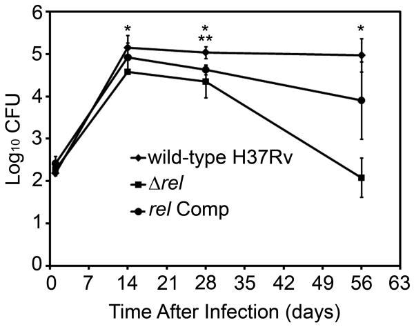
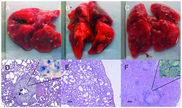
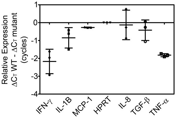
During human latent tuberculosis infection, Mycobacterium tuberculosis likely resides within the nutrient‐starved environment of caseous lung granulomas. The stringent response alarmone (p)ppGpp is synthesized by Rel in response to nutrient starvation, thus enabling tubercle bacilli to restrict growth and shut down metabolism in a coordinated fashion. In this study, we investigated the virulence of a rel‐deficient M. tuberculosis mutant in the guinea pig model. Quantitative reverse‐transcription polymerase chain reaction was used to study the effect of (p)ppGpp deficiency on expression of key cytokine and chemokine genes in guinea pig lungs. The rel‐deficient mutant showed impaired initial growth and survival relative to the wild‐type strain. Loss of Rel was associated with the striking absence of tubercle lesions grossly and of caseous granulomas histologically. The attenuated phenotype of the rel‐deficient mutant was not associated with increased expression of genes encoding the proinflammatory cytokines interferon‐γ and tumor necrosis factor α in the lungs 28 days after infection.


Conflict of interest statement

The authors of this manuscript have no conflicts of interest to report.

Figures

Figure 1
Figure 1
Complementation of Δrel. A. Diagram of expected recombination between integrating plasmid and genomic DNA. B. PCR analysis of genomic DNA. Lanes 2-4 show the presence of rel in wild-type and rel Comp strains. Lanes 6-8 show the presence of a region of the hygromycin gene marking the deletion of rel in both the Δrel and rel Comp strains. Lanes 10-12 show the presence of a region of the kanamycin gene marking the complementation of rel in only the rel Comp strain. C. Diagram of expected sizes of genomic gene fragments expected to bind to probe recognizing region of rel coding sequence. D. Southern blot showing rel as a single copy gene present in the correct fragment size in both wild-type and rel Comp strains.
Figure 2
Figure 2
Reduced initial growth and survival of Δrel strain in the lungs of guinea pigs after low-dose aerosol infection. CFU = colony-forming units. * indicates significant differences between CFU recovered from wild-type and mutant infected lungs (p<=.01). ** indicates the only time point with a significant difference between the wild-type group and the rel Comp group (p=0.004).
Figure 3
Figure 3
Guinea pig lung pathology after 56 days of infection with Mycobacterium tuberculosis. Gross pathology (A-C) and histology (D-F) of lung samples from animals infected with H37Rv (A, D), H37Rv Δrel (B, E), or H37Rv Δrel Comp (C, F). Ziehl-Neelsen stain demonstrating the presence of acid-fast bacilli in the periphery of lung granulomas of animals infected with H37Rv (D inset) and Δrel Comp (F inset). Acid-fast bacilli were not detectable in lung granulomas of Δrel-infected animals (E).
Figure 4
Figure 4
Similar relative abundance of mRNA transcripts from guinea pig lungs following 28 days of infection with Δrel strain compared to wild-type. Data points indicate values from individual Δrel-infected animals. Error bars represent standard deviation. mutant = Δrel strain; WT = wild type (H37Rv) strain; ΔCT = cycle threshold (CT) on Day 28 – CT on Day 1. IFN-γ (p=.06), IL-1B (p=.20), MCP (p=.11), IL-8 (p=.88), TGF-β (p=.47), and TNF-α (p=.003).

Similar articles

Cited by

References

    1. Flynn JL, Chan J. Tuberculosis: latency and reactivation. Infect Immun. 2001;69:4195–201. - PMC - PubMed
    1. Kaplan G, Post FA, Moreira AL, et al. Mycobacterium tuberculosis growth at the cavity surface: a microenvironment with failed immunity. Infect Immun. 2003;71:7099–108. - PMC - PubMed
    1. Grosset J. Mycobacterium tuberculosis in the extracellular compartment: an underestimated adversary. Antimicrob Agents Chemother. 2003;47:833–6. - PMC - PubMed
    1. Nuermberger E, Bishai WR, Grosset JH. Latent tuberculosis infection. Semin Respir Crit Care Med. 2004;25:317–36. - PubMed
    1. Opie ELAJ. Tubercle bacilli in latent tuberculous lesions and in lung tissue without tuberculous lesions. Arch Pathol Lab Med. 1927;4:1–21.

Publication types

MeSH terms