Review
. 2010 Oct;38(5):1349-55.
doi: 10.1042/BST0381349.

Shared paramyxoviral glycoprotein architecture is adapted for diverse attachment strategies

Affiliations
Review

Shared paramyxoviral glycoprotein architecture is adapted for diverse attachment strategies

Thomas A Bowden et al. Biochem Soc Trans. 2010 Oct.

Erratum in

  • Biochem Soc Trans. 2010 Dec;38(6):1723

Abstract

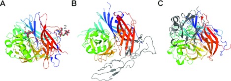
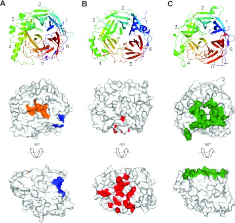
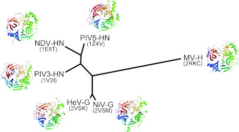
Members within the paramyxovirus subfamily Paramyxovirinae constitute a large number of highly virulent human and animal pathogens. The glycoproteins present on these viruses are responsible for mediating host cell attachment and fusion and are key targets for the design of antiviral entry inhibitors. In the present review, we discuss recent structural studies which have led to a better understanding of the various mechanisms by which different paramyxoviruses use their attachment glycoproteins to hijack specific protein and glycan cell-surface receptors to facilitate viral entry. It is observed that the paramyxovirus attachment glycoprotein consists of a conserved overall structure which includes an N-terminal six-bladed β-propeller domain which is responsible for cell receptor binding. Crystal structures of this domain from different biomedically important paramyxoviruses, including measles, Nipah, Hendra, Newcastle disease and parainfluenza viruses, alone and in complex with their functional cell-surface receptors, demonstrate three contrasting mechanisms of receptor engagement that paramyxoviruses have evolved to confer discreet protein- and glycan-receptor specificity. This structural information highlights the adaptability of the paramyxovirus attachment glycoprotein surface and the potential for the emergence of new and potentially harmful viruses in human hosts.


Figures

Figure 1
Figure 1. Representative structures of the three Paramyxovirinae attachment glycoprotein classes in complex with their human cell-surface receptors
All attachment glycoproteins are superimposed in the same orientation and shown as cartoons coloured as a rainbow with the N-terminus in blue and the C-terminus in red. (A) NDV-HN in complex with DANA at the primary receptor-binding site (1) and thiosialoside at the putative secondary receptor-binding site (2) (PDB code 1USX). DANA and thiosialoside are shown as sticks with carbon atoms coloured grey, oxygen atoms red, nitrogen atoms blue and sulfur atoms yellow. (B) MV-H in complex with the SCR1 and SCR2 domains of CD46 (PDB code 3INB). CD46 is shown as a grey cartoon. (C) NiV-G in complex with the receptor-binding domain of ephrinB2 (PDB code 2VSM). EphrinB2 is shown as a grey cartoon.
Figure 2
Figure 2. Comparison of the receptor-binding sites for HN, H and G Paramyxovirinae attachment glycoproteins
In the top panel, (A) NDV-HN (PDB code 1USX), (B) MV-H (PDB code 3INB) and (C) NiV-G (PDB code 2VSM) are shown as β-propellers and are coloured as in Figure 1. The β-propeller blades are numbered from one to six according to standard nomenclature [10]. In the bottom panels, surface representations for each of the molecules are shown. Cell-surface receptor-binding footprints were calculated using PDBsum [48] and plotted on to the surface of each viral glycoprotein. In (A), the DANA-binding site on NDV-HN is shown in orange and the thiosialoside-binding site is shown in blue. In (B), the CD46-binding site on MV-H is shown in red. In (C), the ephrinB2-binding site is shown in green.
Figure 3
Figure 3. Structural phylogeny of representative receptor-binding β-propeller domains from the Paramyxovirinae
Superpositions were performed using SHP [49] to optimize the probability of equivalence between Cα residue pairs. Following alignment, a pairwise evolutionary distance matrix was constructed and converted into an unrooted tree using PHYLIP [50]. PDB codes are shown in parentheses.

Similar articles

Cited by

References

    1. Lamb R.A., Jardetzky T.S. Structural basis of viral invasion: lessons from paramyxovirus F. Curr. Opin. Struct. Biol. 2007;17:427–436. - PMC - PubMed
    1. Amonsen M., Smith D.F., Cummings R.D., Air G.M. Human parainfluenza viruses hPIV1 and hPIV3 bind oligosaccharides with α2–3-linked sialic acids that are distinct from those bound by H5 avian influenza virus hemagglutinin. J. Virol. 2007;81:8341–8345. - PMC - PubMed
    1. Dorig R.E., Marcil A., Chopra A., Richardson C.D. The human CD46 molecule is a receptor for measles virus (Edmonston strain) Cell. 1993;75:295–305. - PubMed
    1. Naniche D., Varior-Krishnan G., Cervoni F., Wild T.F., Rossi B., Rabourdin-Combe C., Gerlier D. Human membrane cofactor protein (CD46) acts as a cellular receptor for measles virus. J. Virol. 1993;67:6025–6032. - PMC - PubMed
    1. Tatsuo H., Ono N., Yanagi Y. Morbilliviruses use signaling lymphocyte activation molecules (CD150) as cellular receptors. J. Virol. 2001;75:5842–5850. - PMC - PubMed

Publication types