. 2010 Sep 23:5:38.
doi: 10.1186/1750-1326-5-38.

Presenilin-1 regulates induction of hypoxia inducible factor-1α: altered activation by a mutation associated with familial Alzheimer's disease

Affiliations

Presenilin-1 regulates induction of hypoxia inducible factor-1α: altered activation by a mutation associated with familial Alzheimer's disease

Rita De Gasperi et al. Mol Neurodegener. .

Abstract

Background: Mutations in presenilin-1 (Psen1) cause familial Alzheimer's disease (FAD). Both hypoxia and ischemia have been implicated in the pathological cascade that leads to amyloid deposition in AD. Here we investigated whether Psen1 might regulate hypoxic responses by modulating induction of the transcription factor hypoxia inducible factor 1-α (HIF-1α).

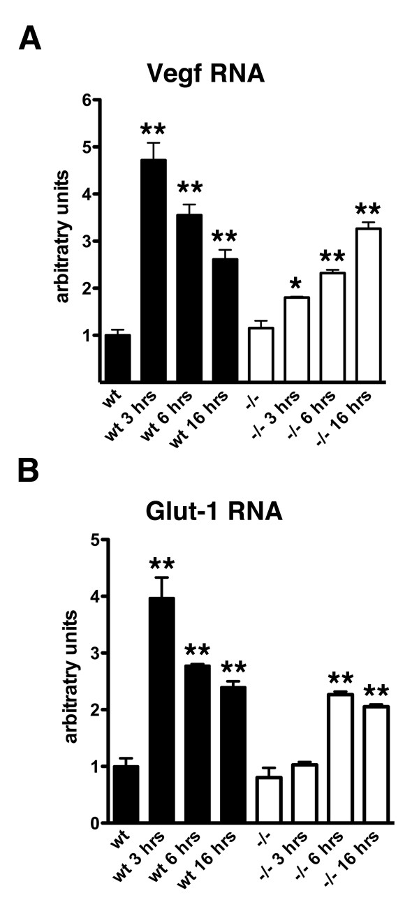
Results: In fibroblasts that lack Psen1 induction of HIF-1α was impaired in response to the hypoxia mimetic cobalt chloride, as well as was induction by insulin and calcium chelation. Reintroduction of human Psen1 using a lentiviral vector partially rescued the responsiveness of Psen1-/- fibroblasts to cobalt chloride induction. HIF-1α induction did not require Psen1's associated γ-secretase activity. In addition, the failure of insulin to induce HIF-1α was not explicable on the basis of failed activation of the phosphatidylinositol 3-kinase (PI3K/Akt) pathway which activated normally in Psen1-/- fibroblasts. Rather we found that basal levels of HIF-1α were lower in Psen1-/- fibroblasts and that the basis for lower constitutive levels of HIF-1α was best explained by accelerated HIF-1α degradation. We further found that Psen1 and HIF-1α physically interact suggesting that Psen1 may protect HIF-1α from degradation through the proteasome. In fibroblasts harboring the M146V Psen1 FAD mutation on a mouse Psen1 null background, metabolic induction of HIF-1α by insulin was impaired but not hypoxic induction by cobalt chloride. Unlike Psen1-/- fibroblasts, basal levels of HIF-1α were normal in FAD mutant fibroblasts but activation of the insulin-receptor pathway was impaired. Interestingly, in Psen1-/- primary neuronal cultures HIF-1α was induced normally in response to cobalt chloride but insulin induction of HIF-1α was impaired even though activation of the PI3K/Akt pathway by insulin proceeded normally in Psen1-/- neuronal cultures. Basal levels of HIF-1α were not significantly different in Psen1-/- neurons and HIF-1α levels were normal in Psen1-/- embryos.

Conclusions: Collectively these studies show that Psen1 regulates induction of HIF-1α although they indicate that cell type specific differences exist in the effect of Psen1 on induction. They also show that the M146V Psen1 FAD mutation impairs metabolic induction of HIF-1α, an observation that may have pathophysiological significance for AD.


Figures

Figure 1
Figure 1
Impaired induction of HIF-1α in Psen1-/- immortalized mouse embryonic fibroblasts. In panel A, the time course of HIF-1α induction in Psen1+/+ (wt) and Psen1-/- (-/-) immortalized mouse embryonic fibroblasts is shown. Cultures were treated with 100 μm cobalt chloride for the indicated times (min). Lysates were prepared and Western blotting was performed using an anti-HIF-1α antibody. The lower panel shows the blot reprobed for β-tubulin. A representative blot is shown from experiments that were performed multiple times. In panels B and C, Psen1+/+ (wt) and Psen1-/- (-/-) cultures were treated with 100 μm cobalt chloride for 4 hours and analyzed for HIF-1α as in (A). The lowest panel in B shows representative samples of wt and -/- fibroblasts probed with the Psen1 antibody 33B10 to confirm the lack of detectible Psen1 expression in Psen1-/- fibroblasts. Panel C shows quantitation of the levels of HIF-1α in data derived from four independent experiments. In panels D and E cultures were serum starved overnight and then treated with insulin for 4 hours after which lysates were prepared and Western blotting performed as in (A). Panel E shows quantitation of the experiment shown in (D). In panel F, Psen1+/+ and Psen1-/- fibroblasts were treated with BAPTA-AM for 1.5 hours. Results from an experiment performed in triplicate is shown. The lower panel shows the blot reprobed for β-tubulin. All panels show representative blots from experiments that were performed multiple times. Western blots for HIF-1α were performed using a rabbit polyclonal antibody. The quantitative results are expressed as the ratio of HIF-1α to β-tubulin and are presented as + SEM. Asterisks in panels C and E indicate values that are different from the corresponding untreated cells (p < 0.05, unpaired t-tests). Other statistical comparisons are discussed in the text.
Figure 2
Figure 2
Reduced activation of HIF-1α downstream target genes in Psen1-/- fibroblasts treated with cobalt chloride. Psen1+/+ (wt) or Psen1-/- fibroblasts were treated with cobalt chloride for the indicated times (hours). Levels of Vegf (A) and Glut-1 (B) RNA were determined by qPCR (n = 9 cultures per group untreated; n = 3 per group treated). (*) indicates p < 0.05 and (**) p < 0.01 vs. untreated control by Dunnett's post-test. Statistical comparisons between groups are discussed further in the text.
Figure 3
Figure 3
Reintroduction of Psen1 into Psen1-/- fibroblasts. Psen1-/- fibroblasts were infected with lentiviruses carrying the human Psen1 cDNA (hPS1-LV) for 24 hours and grown in complete medium for 48 hours. The infected cells were then treated for 4 hours with 100 μM cobalt chloride along with non-infected Psen1-/- cells grown in parallel. The cells were lysed and analyzed for HIF-1α induction by Western blot using anti HIF-1α polyclonal antibodies. Blots were reprobed for anti β-tubulin as a loading control. Psen1 expression was analyzed using the NT.1 mouse monoclonal antibody. FL: full length, NTF: N-terminal fragment. A representative blot is shown in panel A. Panel B shows quantitation of the HIF-1α response (n = 3 samples per group non-infected; n = 4 infected). P values (unpaired t-tests) are indicated for selected comparisons which are discussed further in the text.
Figure 4
Figure 4
Impaired induction of HIF-1α in immortalized mouse embryonic fibroblasts lacking both Psen1 and Psen2. Wild type, Psen1-/- (PS1-/-) and Psen1/Psen2-/- (PS-/-) immortalized fibroblasts were treated with 100 μm cobalt chloride for 4 hours (A) or 2.4 μg/ml of insulin for 6 hours (B). Lysates were prepared and Western blotting was performed using an anti HIF-1α antibody. Lower panels show blots reprobed for β-tubulin. Asterisks (*) indicate lanes that were under loaded based on β-tubulin levels. Note the lowered induction of HIF-1α in the cells lacking both presenilins. Representative blots are shown from experiments that were performed multiple times.
Figure 5
Figure 5
γ-secretase activity is not needed for insulin or cobalt induction of HIF-1α. Psen1+/+ immortalized fibroblasts (A) were treated overnight with the γ-secretase inhibitor XXI and then stimulated with insulin for 6 hours. Western blotting for HIF-1α was performed (upper panel) followed by reprobing of the blot for β-tubulin (middle panel). The same samples were then reblotted and probed for N-cadherin (lower panel). A band for the full length N-cadherin (FL) is indicated. A band that corresponds to the N-cadherin CTF is visible in the γ-secretase inhibitor treated lanes. In panel B, Psen1+/+ fibroblasts were treated overnight with the γ-secretase inhibitor L-685,458 and then stimulated with cobalt chloride for 6 hours. The samples were blotted and probed for HIF-1α (upper panel) and β-actin (middle panel) and then reblotted and probed for N-cadherin (lower panel). In panel C, the experiment in (A) was quantitated with the levels of HIF-1α normalized to β-tubulin. Analysis of the data in panel C (n = 3 per group) revealed significant increases in HIF-1α following treatment with insulin vs. untreated control (p = 0.004, unpaired t-test) or XXI treated vs. insulin + XXI (p = 0.01). There was no difference between insulin treated vs. insulin plus XXI treated cultures (p = 0.29) although XXI treatment alone modestly decreased HIF-1α levels vs. untreated control (p = 0.006). Representative experiments are shown. Asterisks (*) indicate values that are significantly different from corresponding samples that were not treated with insulin.
Figure 6
Figure 6
Normal activation by IGF-1 and insulin of PI3K/Akt in Psen1-/- fibroblasts. Immortalized fibroblast cell lines were treated with IGF-1 (A) or insulin (B) for the indicated times. Western blotting was performed for p-Akt (Ser473) followed by reprobing for total Akt. Panels show representative blots from experiments that were performed multiple times.
Figure 7
Figure 7
Basal levels of HIF-1α are decreased in Psen1-/- fibroblasts. Shown are prolonged exposures of Western blots from non-stimulated Psen1+/+ and Psen1-/- fibroblasts. A representative experiment that was performed multiple times is shown in panel A. In panel B, the experiment in (A) is quantified (asterisk indicates p = 0.0005, unpaired t-test). In panel C, levels of HIF-1α RNA were determined in Psen1 +/+ and Psen1-/- fibroblasts by qPCR (n = 3 cultures per group). Samples were run in triplicate and normalized to the geometric mean of Ppia and Gusb. Asterisk (*) indicates p = 0.0008.
Figure 8
Figure 8
Synthesis rates of HIF-1α are unchanged in Psen1-/- fibroblasts. Psen1+/+ (A) and Psen1-/- (B) fibroblasts were treated with the proteasome inhibitor MG132 for the indicated times (min). Lysates were prepared and Western blotting was performed using an anti-HIF-1α antibody. The lower panels show the blots reprobed for β-tubulin. In panels C and D, the rate of accumulation of HIF-1α normalized to β-tubulin levels from the experiments in panels A and B is shown either as the raw data (C) or normalized to basal levels of HIF-1α in the respective cell types (D). Note that although basal levels of HIF-1α are lower in Psen1-/- fibroblasts, HIF-1α accumulated at similar rates in Psen1+/+ and Psen1-/- fibroblasts. A representative example is shown from experiments that were performed multiple times.
Figure 9
Figure 9
Accelerated degradation of HIF-1α in Psen1-/- fibroblasts. Psen1+/+ and Psen1-/- fibroblasts were treated with cycloheximide. In panel A, a representative experiment is shown with cells treated for the indicated times. Due to the difference in basal HIF-1α levels between Psen1 +/+ and Psen1-/- cells, total protein loaded (15 μg for Psen1+/+ and 30 μg for Psen1 -/-) and blot exposure times were adjusted so that the signal at time 0' would be of similar intensity in Psen1+/+ and Psen1-/- cells to facilitate comparison. In panel B, the % HIF-1α remaining at each time point normalized to β-tubulin is shown. Data is derived from the average of three independently performed experiments.
Figure 10
Figure 10
Physical interaction between HIF-1α and Psen1. Expression vectors carrying no insert, Psen1 or HIF-1α were transfected into HEK 293 cells in the indicated combinations. In panel A, immunoprecipitations (IP) were performed with a polyclonal anti-Psen1 antibody which recognizes the Psen1 NTF or with rabbit IgG as control and Western blotting was performed for HIF-1α. Total extracts without IP (-) (5% of input) were loaded in the two far right lanes (input). Note the co-immunoprecipitation of HIF-1α in the cultures co-transfected with HIF-1α and Psen1. A similar experiment was performed in (B), except that immunoprecipitations were performed with the monoclonal antibody NT.1 which recognizes the Psen1 NTF or with an antibody against the Psen1 loop region. In panel C, immunoprecipitation was done with a monoclonal HIF-1α antibody followed by Western blotting with an antibody against the Psen1 NTF (NT.1) or CTF (33B10). Panels labeled "none" show blots of total extracts prior to immunoprecipitation (input). Bands marked with an asterisk (*) are likely IgGs based on the rate of migration. Panel D shows the co-immunoprecipitation of endogenous HIF-1α by endogenous Psen1. Wild type fibroblasts were treated for 4 hours with 100 μM CoCl2 and 10 μM MG132 to induce HIF-1α accumulation and co-immunoprecipitations were performed as in (A).
Figure 11
Figure 11
Altered activation of HIF-1α in fibroblasts harboring the M146V PSEN1 FAD mutation. Fibroblast cell lines harboring human wild type Psen1 (HuPS1Wt) or the M146V FAD mutant both on the mouse Psen1-/- background were treated with cobalt chloride (A) or insulin (C) as in Figure 1 for the indicated times (hours). Representative blots are shown from experiments that were performed multiple times. The experiment in panel A is quantitated in panel B. In panel D, the insulin induction time course in panel C was quantitated. Asterisks (*) indicate p = 0.04 at 3 hours and p = 0.02 at 6 hours (unpaired t-tests).
Figure 12
Figure 12
Basal levels of HIF-1α are unchanged in M146V FAD mutant fibroblasts. Shown are prolonged exposures of Western blots from the fibroblast lines studied in Figure 11. In panel A, representative blots are shown of samples from individual cultures harboring the human wild type Psen1 (HuPS1Wt) or the M146V FAD mutant. The lower panel shows the blot reprobed for β-tubulin. In panel B, levels of HIF-1α were determined by quantitative Western blotting (n = 5 cultures per genotype) with the levels of HIF-1α normalized to the levels of β-tubulin. Studies were performed both in the presence of serum (FBS) and after overnight serum starvation (-FBS). There were no statistically significant differences between the HuPS1Wt and M146V FAD mutant cell lines.
Figure 13
Figure 13
Decreased insulin-induced activation of the insulin receptor, Akt and GSK-3β in fibroblasts harboring the M146V Psen1 FAD mutation. In panel A, human wild type Psen1 (HuPS1Wt) or M146V FAD mutant cell lines were serum starved overnight and then stimulated with insulin for the indicated times. Western blotting for the phosphorylated IR receptor β (Tyr 1150/1151) or total IR is shown in experiments run in duplicate. The pro-insulin receptor and processed IR are indicated. In panel B, duplicate experiments are shown following Western blotting for p-Akt (Ser473) and total Akt. p-Akt panels are shown at two different exposures. Lower panel shows the blot reprobed for β-tubulin as a loading control. In panel C, the extracts were blotted and probed for p-GSK-3β (Ser9) and total GSK-3β. In panels D, E and F the relative increase in p-IR/total IR (D), p-Akt/total Akt (E) and p-GSK-3β/total GSK-3β (F) are shown for the experiments in A-C.
Figure 14
Figure 14
Altered induction of HIF-1α in Psen1-/- primary neuronal cultures. Primary neuronal cultures were prepared from E15.5-16 embryonic brains and maintained in Neurobasal medium/B27 supplement. Experiments were performed after 5-6 days in vitro. In panel A, cultures were treated with 100 μm cobalt chloride for the indicated times (hrs). In panels B and C, cells were cultured without B27 overnight and then treated with insulin (B) or IGF-1 (C) for six hours. In panel D, cells were cultured without B27 overnight and then treated with IGF-1 for the indicated times. Western blotting for HIF-1α was performed as in Figure 1 and the lower panels show the same blots reprobed for β-tubulin or actin. In panels E and H, the time course of the studies in (A) and (D) are quantitated. The insulin and IGF-1 studies in panels B and C are quantitated in (F) and (G). All panels show representative blots from experiments that were performed multiple times. Asterisks indicate p < 0.05 vs. untreated control (unpaired t-tests).
Figure 15
Figure 15
Activation of HIF-1α downstream target genes in Psen1-/- neurons treated with cobalt chloride or insulin. Levels of Vegf RNA were determined by qPCR following three hours of treatment of wild type or Psen1-/- primary neuronal cultures with cobalt chloride (A, n = 3 cultures per group) or seven hours with insulin (B, n = 2 cultures per group). Samples were run in triplicate and normalized to Ppia. Asterisks indicate p < 0.01 vs. untreated control (unpaired t-tests).
Figure 16
Figure 16
Basal levels of HIF-1α RNA and protein are not significantly changed in Psen1-/- neurons. Basal HIF-1α RNA and protein were measured in neurons in the presence and absence of B27 supplement. In panel A, HIF-1α RNA was measured in neuronal cultures (n = 3/genotype) by qPCR. Samples were run in triplicate and normalized to the geometric means of Ppia and Gusb. In panel B, basal levels of HIF-1α protein were measured in neurons by Western blotting (n = 6 for cultures with B27 and n = 10 for B27 starved cultures). No statistically significant differences were found among the samples (unpaired t-tests). In panel C, representative Western blots are shown of cells cultured with B27 or without (-B27).
Figure 17
Figure 17
Normal activation by insulin of PI3K/Akt in Psen1-/- neurons. Primary neuronal cultures were treated with insulin for the indicated times. In panel A, Western blotting was performed for p-Akt (Ser473) followed by reprobing for total Akt. Shown are representative blots from experiments that were performed multiple times. In panel B, the time course of induction of p-Akt in primary neuronal cultures (n = 2 per genotype per time point) is shown as a ratio of p-Akt/total Akt. There were no significant differences at any time point (unpaired t-tests).
Figure 18
Figure 18
No change in HIF-1α levels in Psen1-/- embryonic brain. HIF-1α levels were determined by Western blotting of extracts from E14.5 (A) or E18.5 (B) brain from wild type (wt) or homozygous (-/-) Psen1 mutant mice. Representative experiments are shown. Lower panels show the same blots reprobed for β-tubulin. HIF-1α levels were quantitated as a ratio to β-tubulin at E14.5 (C) and E18.5 (D) (n = 4 wild type and 2 Psen1-/- at each age). There were no differences between wild type (wt) and Psen1-/- at either age (unpaired t-tests).

Similar articles

Cited by

References

    1. Selkoe DJ. Alzheimer's disease: genes, proteins, and therapy. Physiol Rev. 2001;81(2):741–766. - PubMed
    1. Ertekin-Taner N. Genetics of Alzheimer's disease: a centennial review. Neurol Clin. 2007;25(3):611–667. doi: 10.1016/j.ncl.2007.03.009. - DOI - PMC - PubMed
    1. Alzheimer Disease & Frontotemporal Dementia Mutation Database. http://www.molgen.ua.ac.be/ADMutations
    1. Lleo A, Berezovska O, Growdon JH, Hyman BT. Clinical, pathological, and biochemical spectrum of Alzheimer disease associated with PS-1 mutations. Am J Geriatr Psychiatry. 2004;12(2):146–156. - PubMed
    1. Walsh DM, Selkoe DJ. A beta oligomers - a decade of discovery. J Neurochem. 2007;101(5):1172–1184. doi: 10.1111/j.1471-4159.2006.04426.x. - DOI - PubMed

LinkOut - more resources