. 2010 Sep 23:7:77.
doi: 10.1186/1742-4690-7-77.

Self-association of the Lentivirus protein, Nef

Affiliations

Self-association of the Lentivirus protein, Nef

Youn Tae Kwak et al. Retrovirology. .

Abstract

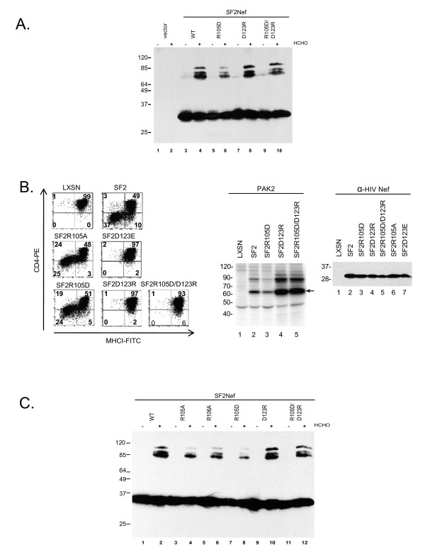
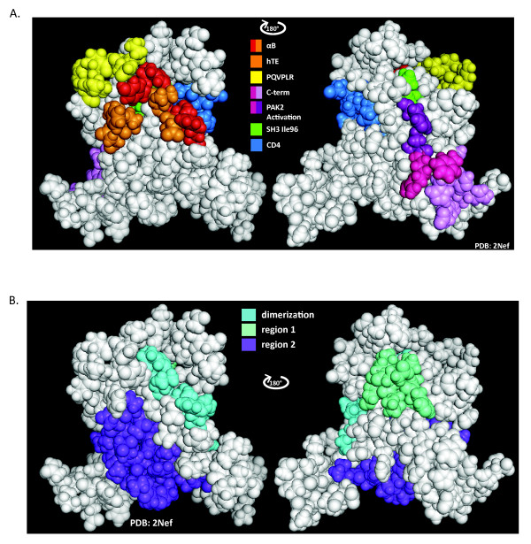
Background: The HIV-1 pathogenic factor, Nef, is a multifunctional protein present in the cytosol and on membranes of infected cells. It has been proposed that a spatial and temporal regulation of the conformation of Nef sequentially matches Nef's multiple functions to the process of virion production. Further, it has been suggested that dimerization is required for multiple Nef activities. A dimerization interface has been proposed based on intermolecular contacts between Nefs within hexagonal Nef/FynSH3 crystals. The proposed dimerization interface consists of the hydrophobic B-helix and flanking salt bridges between R105 and D123. Here, we test whether Nef self-association is mediated by this interface and address the overall significance of oligomerization.

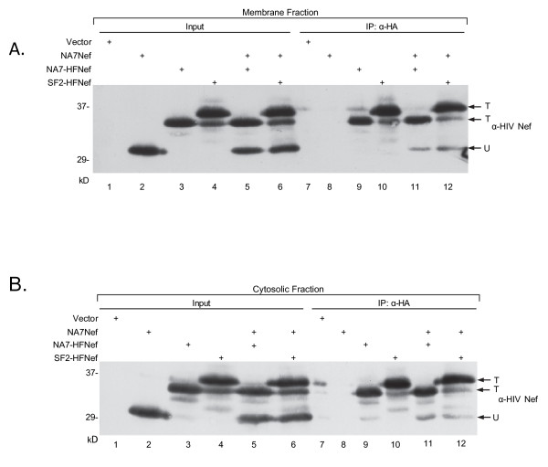
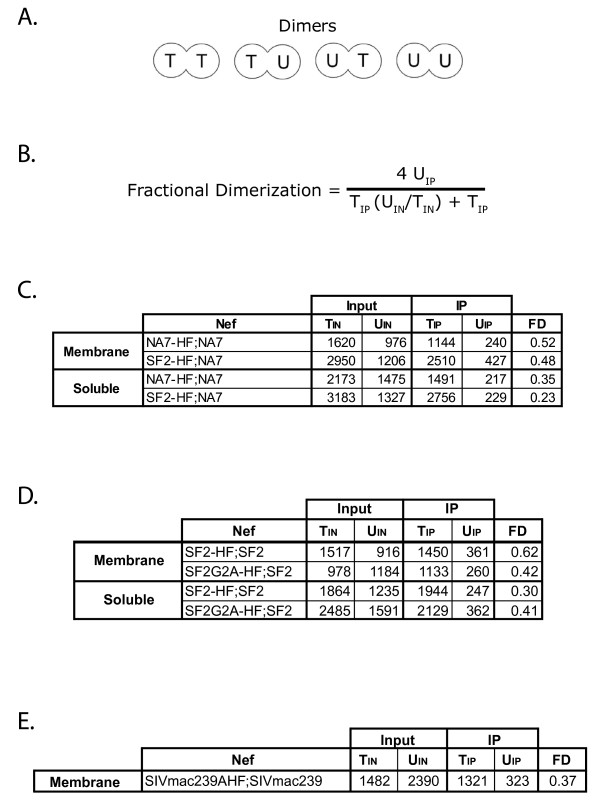
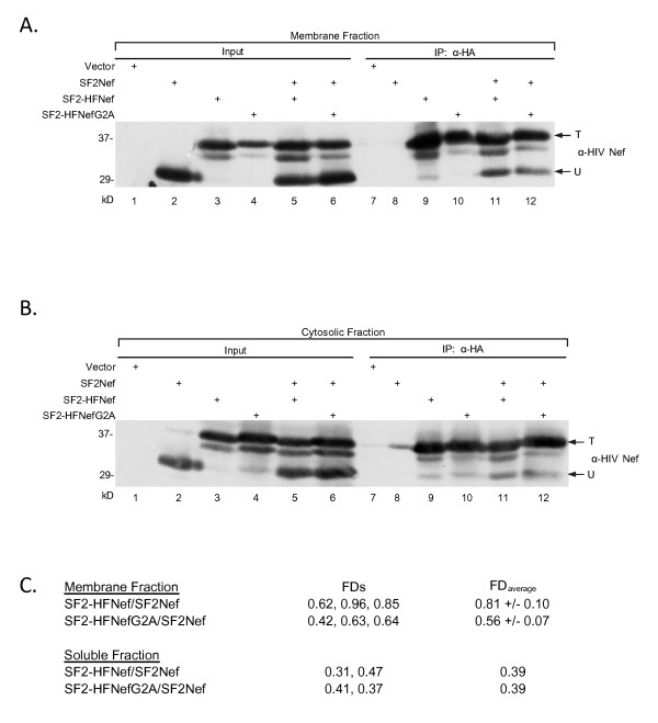
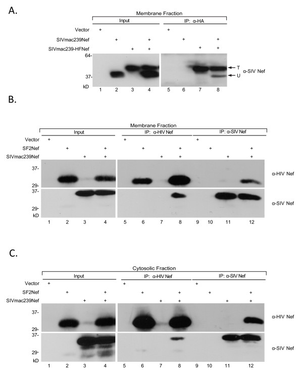
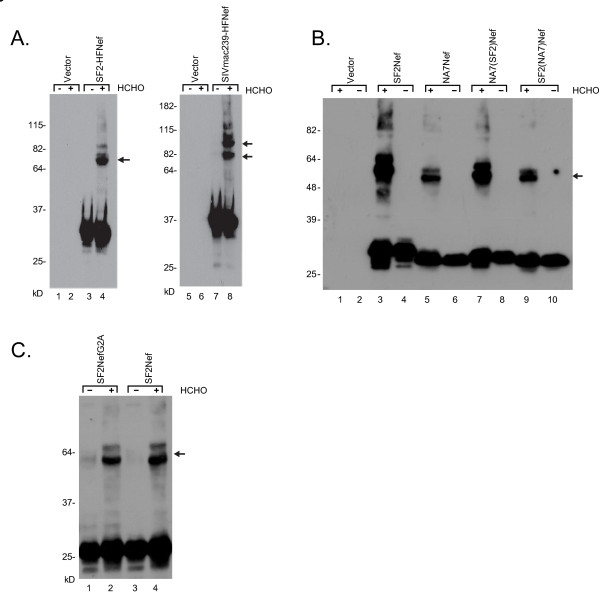
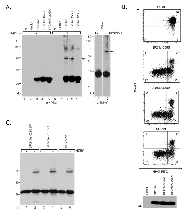
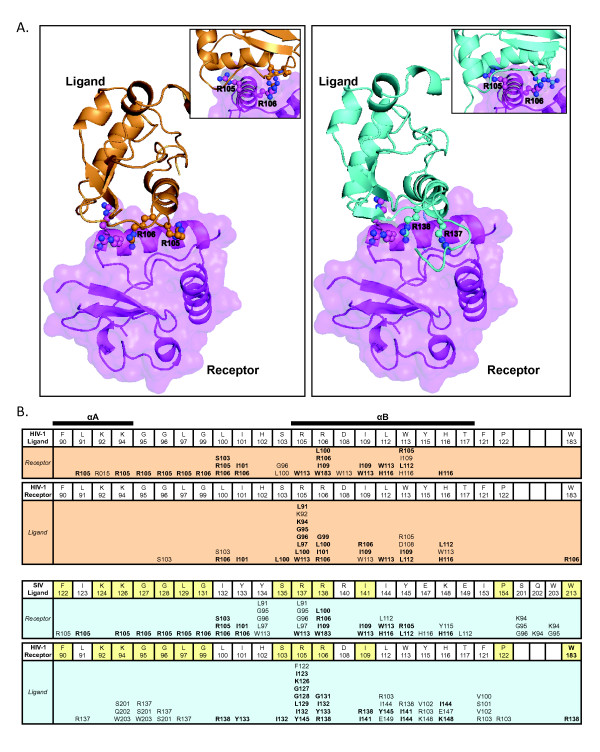
Results: By co-immunoprecipitation assays, we demonstrated that HIV-1Nef exists as monomers and oligomers with about half of the Nef protomers oligomerized. Nef oligomers were found to be present in the cytosol and on membranes. Removal of the myristate did not enhance the oligomerization of soluble Nef. Also, SIVNef oligomerizes despite lacking a dimerization interface functionally homologous to that proposed for HIV-1Nef. Moreover, HIV-1Nef and SIVNef form hetero-oligomers demonstrating the existence of homologous oligomerization interfaces that are distinct from that previously proposed (R105-D123). Intracellular cross-linking by formaldehyde confirmed that SF2Nef dimers are present in intact cells, but surprisingly self-association was dependent on R105, but not D123. SIV(MAC239)Nef can be cross-linked at its only cysteine, C55, and SF2Nef is also cross-linked, but at C206 instead of C55, suggesting that Nefs exhibit multiple dimeric structures. ClusPro dimerization analysis of HIV-1Nef homodimers and HIV-1Nef/SIVNef heterodimers identified a new potential dimerization interface, including a dibasic motif at R105-R106 and a six amino acid hydrophobic surface.

Conclusions: We have demonstrated significant levels of intracellular Nef oligomers by immunoprecipitation from cellular extracts. However, our results are contrary to the identification of salt bridges between R105 and D123 as necessary for self-association. Importantly, binding between HIV-1Nef and SIVNef demonstrates evolutionary conservation and therefore significant function(s) for oligomerization. Based on modeling studies of Nef self-association, we propose a new dimerization interface. Finally, our findings support a stochastic model of Nef function with a dispersed intracellular distribution of Nef oligomers.


Figures

Figure 1
Figure 1
Assays for Nef oligomers by immunoprecipitation. (A) 293T cells were transfected to express untagged NA7Nef (lower molecular weight band, lane 2) and tagged NA7-HFNef and SF2-HFNef (higher molecular weight bands, lanes 3 and 4) singly and in the combinations of a tagged Nef with untagged NA7Nef. The membrane-associated protein fraction was prepared and Nef expression was determined by Western blot analysis (Input, lanes 1-6). Immunoprecipitations were also performed with anti-HA monoclonal antibody and were analyzed by SDS/PAGE and Western blot analysis (IP: α-HA, lanes 7-12). Antibody for the Western blots was sheep anti-HIV-1Nef. Arrows, untagged NA7Nef (U) present as hetero-oligomer and total tagged Nef (T). Upper T for SF2-HFNef and lower T for NA7-HFNef. (B) Same as in (A) with the corresponding cytoplasmic protein fractions. Note that in (A) the slightly lower molecular weight band present in the SF2-HFNef lanes running the same distance as the NA7-HFNef appears to have been proteolytically modified. In the soluble fractions additional minor proteolytic fragments are present.
Figure 2
Figure 2
Estimation of Fractional Dimerization. (A) There are three forms of Nef dimers possible- homodimers of tagged Nefs (T2) and untagged (U2) Nefs and heterodimers (TU, UT). We have assumed a random assortment of tagged and untagged Nefs with a binomial distribution (1:2:1) of dimeric forms. (B) The equation for the fractional dimerization (FD). (C) The corrected densities (divided by 1000), determined by ImageJ, for the experiment presented in Figure 1A, B were used to calculate FD's. (D) The corrected densities/1000 determined by ImageJ for the experiment presented in Figure 3A, B and the calculated FD's are given. (E) The corrected densities/1000 for the experiment in Figure 4A and the FD are presented.
Figure 3
Figure 3
Myristoylation defective Nef does not exhibit enhanced oligomerization. (A, B) The abilities of untagged SF2Nef to associate with tagged SF2-HFNef or SF2-HFNefG2A in membrane and soluble fractions were compared using the immunoprecipitation assay. The co-immunoprecipitated SF2Nef is indicated by an arrow marked "U" to the right of lane 12 (lanes 11 and 12). The total tagged Nef is indicated by an arrow marked ''T''. (A), Membrane-bound Nef. (B), Cytosolic Nef. (C) Data from additional experiments were combined to assess the FDs for the binding of SF2Nef to SF2-HFNef and SF2-HFNefG2A. Analysis was performed as in Figure 2.
Figure 4
Figure 4
SIVMAC239Nef oligomerizes and also forms heterologous oligomers with SF2Nef. (A) 293T cells were transfected to express tagged SIVMAC239-HFNef and untagged SIVMAC239Nef, singly and in combination. Membrane protein fractions were prepared and Nef expression was determined by Western blot analysis (Input). Immunoprecipitations were performed with anti-HA monoclonal antibody and the immunoprecipitates analyzed by SDS/PAGE followed by Western blotting with monoclonal anti-SIV antibody (IP:α-HA). Arrows, total tagged Nef (T) and untagged Nef bound to tagged Nef (U). (B and C) 293T cells were transfected to express SF2Nef and SIVMAC239Nef singly and in combination. Membrane (B) and soluble (C) protein fractions were prepared and Nef expression determined by Western blot analysis with anti-HIVNef monoclonal antibody (Input, upper panels) and anti-SIVNef monoclonal antibody (Input, lower panels). The membrane and soluble fractions were also immunoprecipitated with polyclonal anti-HIV-1Nef (IP: α-HIV-1Nef) and polyclonal anti-SIVNef (IP: α-SIV Nef). The immunoprecipitates were analyzed by Western blot with monoclonal anti-HIV Nef (upper panels) and monoclonal anti-SIVNef (lower panels).
Figure 5
Figure 5
Cross-linking Nef dimers in intact cells with formaldehyde. (A) 293T cells were transfected to express SF2-HFNef or SIVMAC239-HFNef. Intact cells were incubated with 0.5% formaldehyde for 10 minutes at room temperature. Clarified whole cell lysates were prepared and Nef detected by Western blot analysis. (-), incubation without formaldehyde. (+), incubation with formaldehyde. Arrows, cross-linked Nef dimers. (B) Assay of intracellular oligomerization with NA7Nef/SF2Nef chimeras. The chimeric expression vectors were made by substituting coding sequence for amino acids 24-200 SF2Nef into NA7Nef and the same coding sequence for NA7Nef into SF2Nef. Oligomerization was assayed by formaldehyde cross-linking in intact cells as in (A). NA7(SF2)Nef, NA7Nef with coding sequence for amino acids 24-200 replaced by coding sequence for the same amino acids from SF2Nef. SF2(NA7)Nef, the reciprocal construct as NA7(SF2)Nef. (-), incubation without formaldehyde; (+), incubation with formaldehyde. Arrow, cross-linked Nef dimer. (C), The ability of SF2NefG2A to associate with itself was compared to SF2Nef by the formaldehyde cross-linking assay with intact cells. Arrow to the right of lane 4 indicates cross-linked Nef (lanes 2 and 4). (+), Cells incubated with 0.5% formaldehyde for 10 minutes at room temperature. (-), Cells incubated in the absence of formaldehyde.
Figure 6
Figure 6
Cross-linking Nefs dimers with BM[PEG]3. (A) 293T cells were transfected to express SF2Nef, SF2NefC55S and SF2NefC206X (left, lanes 1-10), and SIVMAC239Nef (right, lanes 11 and 12). Clarified whole cell Iysates were incubated with BM[PEG]3. Nef was then detected by Western blot analysis. (-), clarified whole cell Iysates incubated in the absence of BM[PEG]3 for 1 hour at room temperature (lanes 1-5 and 11). (+), clarified whole cell Iysates incubated in the presence of 0.5 mM BM[PEG]3 for 1 hour at room temperature (lanes 6-10 and 12). The presence of crosslinked Nefs was detected by Western Blot analysis. NT, 293T cells not transfected. (B) SF2Nef, SF2C55S, and SF2C206X were assayed for CD4 downregulation and MHCI downregulation in transduced CEM cells (top). The level of expression of SF2Nef, SF2NefC55S and SF2NefC206X in CEM cells was determined by Western blot analysis (bottom). (C) Lysates from transfected 293T cells expressing SF2NefC206X, SF2NefC55S, and SF2Nef were cross-linked by 0.5% formaldehyde.
Figure 7
Figure 7
R105 and R106 are critical for Nef oligomerization. (A), 293T cells were transfected to express SF2-HFNef (WT), SF2-HFNefR105D (R105D), SF2-HFNefD123R (D123R), and SF2-HFNefR105D/D123R (R105D/D123R). The presence of Nef dimers was detected by treatment of intact cells with 0.5% formaldehyde (+). No formaldehyde (-). (B) Transduced CEM cells expressing SF2Nef, SF2NefR105A, SF2NefD123E, SF2NefR105D, SF2NefD123R, or SF2NefR105D/D123R plus the vector (LXSN) positive control were assayed for CD4 and MHCI cell surface expression by flow cytometry analysis (left) , PAK2 activation (middle), and level of Nef expression by Western blot analysis (right). (C) Role of R105 and R106 in dimerization was investigated as in (A). SF2-HFNefR105A (R105A), SF2-HFNefR106A (R106A).
Figure 8
Figure 8
Three dimensional models of HIV-1 Nef homodimer and HIV-1 Nef/SIVMAC239Nef heterodimer. (A) Three dimensional representation of HIV-1Nef interacting with itself (left) and SIVMAC239Nef (right), identified by rigid body docking using ClusPro. In both docking experiments, HIV-1Nef (PDB 1AVZ, Chain A) was entered as receptor (left and right- magenta cartoon and transparent surface). In the homodimerization docking, the same Nef was entered as ligand (left, light orange cartoon). In the heterodimerization docking SIVMAC239Nef (PDB 3IK5, Chain A) was entered as ligand (right, cyan cartoon). The models shown here represent the two complexes generated by ClusPro in which SIVMAC239Nef and HIV-1Nef interact with the receptor HIV-1Nef in a nearly identical manner. R105 and R106 for HIV-1Nef and SIVMAC239Nef R137 and R138 are represented as ball and stick. Inserts show receptor R105 inserted into a pocket of ligand Nef. (B) Residues interacting in the interface of the HIV-1Nef homodimer, and residues interacting in the interface of the HIV-1Nef/SIVMAC239Nef heterodimer presented in (A). The three rows of amino acids from F90 to W183 for HIV-1Nef and F122 to W213 for SIVMAC239Nef contain all of the residues present at the two dimer interfaces. In the case of SIVMAC239Nef, the residues are orthologs of HIV-1Nef residues. In the heterodimer section (lower half), the HIV-1Nef and SIVMAC239Nef residues that are identical between the two proteins are shaded yellow. Under each boxed residue are the residue or residues that interact with the designated amino acid. Identical interactions between the homodimer and the heterodimer are in bold font. Amino acids S201, Q202, and W203 in SIVMAC239Nef correspond to HIV-1Nef internal loop amino acids 171-173 which are deleted in the HIV-1Nef core construct. The extents of conservation at each amino acid position presented in (B) for HIV-1 subtype B Nefs are: F90, 99%; L91, 98%; K92, 98% for R or K; K94, 95%; G95, 99%; G96, 99%; L97, 99%; G99, 99%; L100, 99% for L, I, or M; I101, 98% for I or V; S103, 98%; R105, 99% for K, R, or Q; R106, 99%; D108, 99% for D or E; I109, 99%; L112, 99%; W113, 99%; Y115, 97%; H116, 99% for H or N; T117, 99%; F121, 99%; P122, 99%; W183, 99%. All positions with multiple residues only have amino acids that have positive BLOSUM62 scores (53).
Figure 9
Figure 9
Three dimensional representations of the functional regions of Nef that interact with host cell proteins or represent putative dimerization domains. (A) left, Red and Orange, αB and flanking loop amino acids of the Nef/Nef interface in hexagonal crystals of PDB 1AVZ (amino acids D108, L112, Y115, H116, F121, P122, D123); Orange, residues that interact with human thioesterase (amino acids D108, L112, F121, P122, D123); Yellow, PQVPLR in the proline helix of the Nef SH3 binding domain (amino acids 72-77); Blue, residues that interact with the cytoplasmic tail of CD4 (amino acids W57, L58, E59, G95, L97, R106, L110); right, Image is rotated 180 degrees. Lilac and Magenta, C-terminal Nef/Nef interface seen in cubic crystals of PDB 1AVV (amino acids F139, R188, F191, H192, H193, R196, E197); Magenta and Purple, residues known to play a role in PAK-2 activation (amino acids H89, S187, R188, F191); Green, residues that form a hydrophobic pocket interacting with Ile96 of the RT loop of the Hck SH3 domain (amino acids L87, F90, W113, I114). (B) left, Cyan, critical residues identified in the protein-protein docking experiments, showcased in Figure 8. Only residues that interact in both the receptor and ligand in both the homodimeric and heterodimeric models are indicated (amino acids L100, I101, R105, R106, I109, L112, W113, H116); blue violet, one of two regions (region 2) of HIV-1Nef that is highly conserved in SIV Nef with 22 out of 27 identities (amino acids 122 to 148). right, Seafoam green, second of two regions (region 1) of HIV-1 Nef that is highly conserved in SIV Nef with 11 out of 12 identities (amino acids 88-99).

Similar articles

Cited by

References

    1. Foster JL, Garcia JV. Role of Nef in HIV-1 replication and pathogenesis. Adv Pharmacol. 2007;55:389–409. full_text. - PubMed
    1. Kirchhoff F, Schindler M, Specht A, Arhel N, Munch J. Role of Nef in primate lentiviral immunopathogenesis. Cell Mol Life Sci. 2008;65:2621–2636. doi: 10.1007/s00018-008-8094-2. - DOI - PMC - PubMed
    1. Kestler HW, Ringler DJ, Mori K. et al.Importance of the nef gene for maintenance of high virus loads and for development of AIDS. Cell. 1991;65:651–662. doi: 10.1016/0092-8674(91)90097-I. - DOI - PubMed
    1. Gorry PR, McPhee DA, Verity E, Dyer WB, Wesselingh SL, Learmont J, Sullivan JS, Roche M, Zaunders JJ, Gabuzda D, Crowe SM, Mills J, Lewin SR, Brew BJ, Cunningham AL, Churchill MJ. Pathogenicity and immunogenicity of attenuated, nef-deleted HIV-1 strains in vivo. Retrovirology. 2007;4:66. doi: 10.1186/1742-4690-4-66. - DOI - PMC - PubMed
    1. Arold S, Franken P, Strub MP, Hoh F, Benichou S, Benarous R, Dumas C. The crystal structure of HIV-1 Nef protein bound to the Fyn kinase SH3 domain suggests a role for this complex in altered T cell receptor signaling. Structure. 1997;5(10):1361–1372. doi: 10.1016/S0969-2126(97)00286-4. - DOI - PubMed

Publication types

MeSH terms

Substances

LinkOut - more resources