

Identification of NCAM that interacts with the PHE-CoV spike protein
- PMID: 20863409
- PMCID: PMC2955716
- DOI: 10.1186/1743-422X-7-254
Identification of NCAM that interacts with the PHE-CoV spike protein
Abstract
Background: The spike proteins of coronaviruses associate with cellular molecules to mediate infection of their target cells. The characterization of cellular proteins required for virus infection is essential for understanding viral life cycles and may provide cellular targets for antiviral therapies.
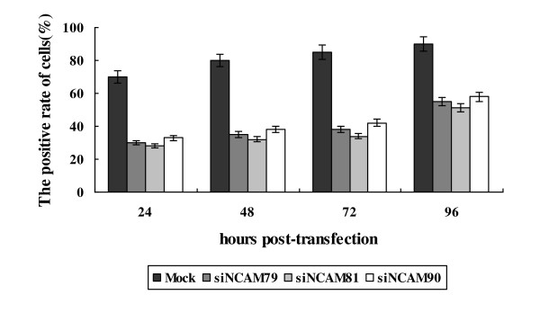
Results: We identified Neural Cell Adhesion Molecule (NCAM) as a novel interacting partner of the PHE-CoV S protein. A T7 phage display cDNA library from N2a cells was constructed, and the library was screened with the soluble PHE-CoV S glycoproteins. We used a coimmunoprecipitation assay to show that only the NCAM was a binding partner of spike protein. We found that a soluble form of anti-NCAM antibody blocked association of the PHE-CoV with N2a cells. Furthermore, double-stranded siRNA targeted against NCAM inhibited PHE-CoV infection.
Conclusions: A novel interaction was identified between NCAM and spike protein and this association is critical during PHE-CoV infection.
Figures


Similar articles
-
A small region of porcine hemagglutinating encephalomyelitis virus spike protein interacts with the neural cell adhesion molecule.Intervirology. 2015;58(2):130-7. doi: 10.1159/000381060. Epub 2015 Apr 25. Intervirology. 2015. PMID: 25925196 Free PMC article.
-
In vitro inhibition of porcine hemagglutinating encephalomyelitis virus replication with siRNAs targeting the spike glycoprotein and replicase polyprotein genes.Intervirology. 2012;55(1):53-61. doi: 10.1159/000323523. Epub 2011 Mar 3. Intervirology. 2012. PMID: 21372550 Free PMC article.
-
Highly conserved regions within the spike proteins of human coronaviruses 229E and NL63 determine recognition of their respective cellular receptors.J Virol. 2006 Sep;80(17):8639-52. doi: 10.1128/JVI.00560-06. J Virol. 2006. PMID: 16912312 Free PMC article.
-
Coronavirus spike proteins in viral entry and pathogenesis.Virology. 2001 Jan 20;279(2):371-4. doi: 10.1006/viro.2000.0757. Virology. 2001. PMID: 11162792 Free PMC article. Review. No abstract available.
-
[Cell entry mechanisms of coronaviruses].Uirusu. 2009 Dec;59(2):215-22. doi: 10.2222/jsv.59.215. Uirusu. 2009. PMID: 20218330 Review. Japanese.
Cited by
-
Development and evaluation of an immunochromatographic strip for rapid detection of porcine hemagglutinating encephalomyelitis virus.Virol J. 2012 Aug 24;9:172. doi: 10.1186/1743-422X-9-172. Virol J. 2012. PMID: 22920192 Free PMC article.
-
Neurological manifestations of coronavirus infections, before and after COVID-19: a review of animal studies.J Neurovirol. 2021 Dec;27(6):864-884. doi: 10.1007/s13365-021-01014-7. Epub 2021 Nov 2. J Neurovirol. 2021. PMID: 34727365 Free PMC article. Review.
-
Genetic diversity and evolution of porcine hemagglutinating encephalomyelitis virus in Guangxi province of China during 2021-2024.Front Microbiol. 2024 Oct 9;15:1474552. doi: 10.3389/fmicb.2024.1474552. eCollection 2024. Front Microbiol. 2024. PMID: 39444682 Free PMC article.
-
Porcine Hemagglutinating Encephalomyelitis Virus: A Review.Front Vet Sci. 2019 Feb 27;6:53. doi: 10.3389/fvets.2019.00053. eCollection 2019. Front Vet Sci. 2019. PMID: 30873421 Free PMC article. Review.
-
Phage Display Technique as a Tool for Diagnosis and Antibody Selection for Coronaviruses.Curr Microbiol. 2021 Apr;78(4):1124-1134. doi: 10.1007/s00284-021-02398-9. Epub 2021 Mar 9. Curr Microbiol. 2021. PMID: 33687511 Free PMC article. Review.
References
-
- Vijgen L, Keyaerts E, Lemey P, Maes P, Van Reeth K, Nauwynck H, Pensaert M, Van Ranst M. Evolutionary history of the closely related group 2 coronaviruses: porcine hemagglutinating encephalomyelitis virus, bovine coronavirus, and human coronavirus OC43. J Virol. 2006;80:7270–7274. doi: 10.1128/JVI.02675-05. - DOI - PMC - PubMed
-
- Miura TA, Travanty EA, Oko L, Bielefeldt-Ohmann H, Weiss SR, Beauchemin N, Holmes KV. The spike glycoprotein of murine coronavirus MHV-JHM mediates receptor-independent infection and spread in the central nervous systems of Ceacam1a(-/-) mice. Journal of Virology. 2008;82:755–763. doi: 10.1128/JVI.01851-07. - DOI - PMC - PubMed
Publication types
MeSH terms
Substances
LinkOut - more resources
Full Text Sources
Research Materials
Miscellaneous
