

Are normative values for LV geometry and mass based on fundamental imaging valid with use of harmonic imaging?
- PMID: 20863657
- PMCID: PMC2993783
- DOI: 10.1016/j.echo.2010.08.014
Are normative values for LV geometry and mass based on fundamental imaging valid with use of harmonic imaging?
Abstract
Background: Multiple studies have reported echocardiographically determined normal reference values for left ventricular (LV) mass (LVM) derived using fundamental imaging (FI). Modern ultrasound systems now use harmonic imaging (HI) because of the improved LV endomyocardial definition. However, the 2005 American Society of Echocardiography (ASE) recommendations noted that the applicability of the reference values to HI-derived measurements has not been established.
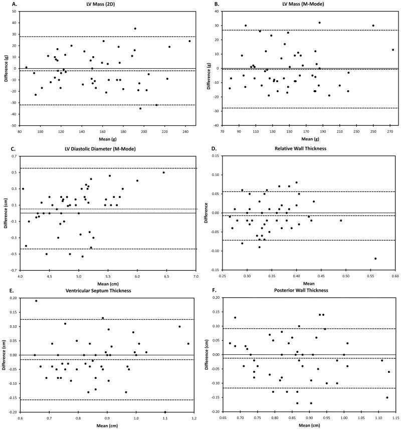
Methods: LVM and LV end-diastolic volume, diameter, and wall thickness were determined using HI in healthy subjects (n = 251), including normal-weight (NW) (body mass index < 25 kg/m², n = 149, 68% women) and otherwise healthy, overweight (OW) (body mass index ≥ 25 and <30 kg/m², n = 102, 41% women) groups. Measurements were compared with ASE-endorsed reference values. The agreement between FI and HI was determined in a prospective cohort of 51 subjects.
Results: Two-dimensional (2D) derived LV volumes were similar between NW and OW subjects, although M-mode (MM)-derived LV diameters were slightly greater in OW subjects. 2D and MM-derived LVM was greater in OW compared with NW subjects, including after adjustment by height or height²·⁷; however, indexing to body surface area eliminated these differences. The partition values for abnormal 2D and MM-derived LVM were generally greater in NW and OW subjects of both sexes compared with the ASE-endorsed values (except MM-derived LVM in NW men). However, there were no significant differences in LVM determined by HI compared with FI in a prospectively studied cohort.
Conclusions: Reference values for LVM derived from NW and OW cohorts are generally higher than the ASE-endorsed referenced values. The difference between NW and ASE-endorsed values is unlikely to result from the use of HI rather than FI, because there is excellent agreement between these two imaging modalities. This study emphasizes the need to update normal reference values to reflect modern imaging methods.
Copyright © 2010 American Society of Echocardiography. Published by Mosby, Inc. All rights reserved.
Figures
Similar articles
-
LV mass assessed by echocardiography and CMR, cardiovascular outcomes, and medical practice.JACC Cardiovasc Imaging. 2012 Aug;5(8):837-48. doi: 10.1016/j.jcmg.2012.06.003. JACC Cardiovasc Imaging. 2012. PMID: 22897998 Free PMC article. Review.
-
Normative Left Ventricular M-Mode Echocardiographic Values in Preterm Infants up to 2 kg.J Am Soc Echocardiogr. 2017 Aug;30(8):781-789.e4. doi: 10.1016/j.echo.2017.04.010. Epub 2017 Jun 7. J Am Soc Echocardiogr. 2017. PMID: 28599830 Free PMC article.
-
How to calculate left ventricular mass in routine practice? An echocardiographic versus cardiac magnetic resonance study.Arch Cardiovasc Dis. 2011 May;104(5):343-51. doi: 10.1016/j.acvd.2011.04.003. Epub 2011 Jun 14. Arch Cardiovasc Dis. 2011. PMID: 21693371
-
Comparison between fundamental and second-harmonic imaging echocardiography for calculation of left ventricular mass in children.Clin Physiol Funct Imaging. 2005 Jul;25(4):223-5. doi: 10.1111/j.1475-097X.2005.00612.x. Clin Physiol Funct Imaging. 2005. PMID: 15972024 Clinical Trial.
-
Reference Values for Real Time Three-Dimensional Echocardiography-Derived Left Ventricular Volumes and Ejection Fraction: Review and Meta-Analysis of Currently Available Studies.Echocardiography. 2015 Dec;32(12):1841-50. doi: 10.1111/echo.12972. Epub 2015 Jun 6. Echocardiography. 2015. PMID: 26053260 Review.
Cited by
-
LV mass assessed by echocardiography and CMR, cardiovascular outcomes, and medical practice.JACC Cardiovasc Imaging. 2012 Aug;5(8):837-48. doi: 10.1016/j.jcmg.2012.06.003. JACC Cardiovasc Imaging. 2012. PMID: 22897998 Free PMC article. Review.
-
Genetic association of left ventricular mass assessed by M-mode and two-dimensional echocardiography.J Hypertens. 2016 Jan;34(1):88-96. doi: 10.1097/HJH.0000000000000765. J Hypertens. 2016. PMID: 26556563 Free PMC article.
-
Longitudinal determinants of left ventricular mass and geometry: the Coronary Artery Risk Development in Young Adults (CARDIA) Study.Circ Cardiovasc Imaging. 2013 Sep;6(5):769-75. doi: 10.1161/CIRCIMAGING.112.000450. Epub 2013 Aug 6. Circ Cardiovasc Imaging. 2013. PMID: 23922005 Free PMC article.
References
-
- Ganau A, Devereux RB, Roman MJ, de Simone G, Pickering TG, Saba PS, et al. Patterns of left ventricular hypertrophy and geometric remodeling in essential hypertension. J Am Coll Cardiol. 1992;19:1550. - PubMed
-
- de Simone G, Devereux RB, Roman MJ, Alderman MH, Laragh JH. Relation of obesity and gender to left ventricular hypertrophy in normotensive and hypertensive adults. Hypertension. 1994;23:600–6. - PubMed
-
- Lauer MS, Anderson KM, Larson MG, Levy D. A new method for indexing left ventricular mass for differences in body size. Am J Cardiol. 1994;74:487–91. - PubMed
-
- Shub C, Klein AL, Zachariah PK, Bailey KR, Tajik AJ. Determination of left ventricular mass by echocardiography in a normal population: effect of age and sex in addition to body size. Mayo Clin Proc. 1994;69:205–11. - PubMed
-
- Ilercil A, O’Grady MJ, Roman MJ, Paranicas M, Lee ET, Welty TK, et al. Reference values for echocardiographic measurements in urban and rural populations of differing ethnicity: the Strong Heart Study. J Am Soc Echocardiogr. 2001;14:601–11. - PubMed
Publication types
MeSH terms
Grants and funding
- R01HL071782/HL/NHLBI NIH HHS/United States
- R01 HL071782/HL/NHLBI NIH HHS/United States
- R21HL094668/HL/NHLBI NIH HHS/United States
- S10RR14778/RR/NCRR NIH HHS/United States
- R21 HL094668/HL/NHLBI NIH HHS/United States
- KL2RR024994/RR/NCRR NIH HHS/United States
- UL1 TR000448/TR/NCATS NIH HHS/United States
- KL2 RR024994/RR/NCRR NIH HHS/United States
- KL2 TR000450/TR/NCATS NIH HHS/United States
- K12 RR023249/RR/NCRR NIH HHS/United States
- K12RR023249/RR/NCRR NIH HHS/United States
- UL1RR024992/RR/NCRR NIH HHS/United States
- UL1 RR024992/RR/NCRR NIH HHS/United States
- S10 RR014778/RR/NCRR NIH HHS/United States
- L30 HL074749/HL/NHLBI NIH HHS/United States
LinkOut - more resources
Full Text Sources
